 Spanish
  Chinese
  Russian
  German
  French
  Japanese
  Portuguese
  Hindi
Hindi Our Group organises 3000+ Global Conferenceseries Events every year across USA, Europe & Asia with support from 1000 more scientific Societies and Publishes 700+ Open Access Journals which contains over 50000 eminent personalities, reputed scientists as editorial board members.
Open Access Journals gaining more Readers and Citations
700 Journals and 15,000,000 Readers Each Journal is getting 25,000+ Readers
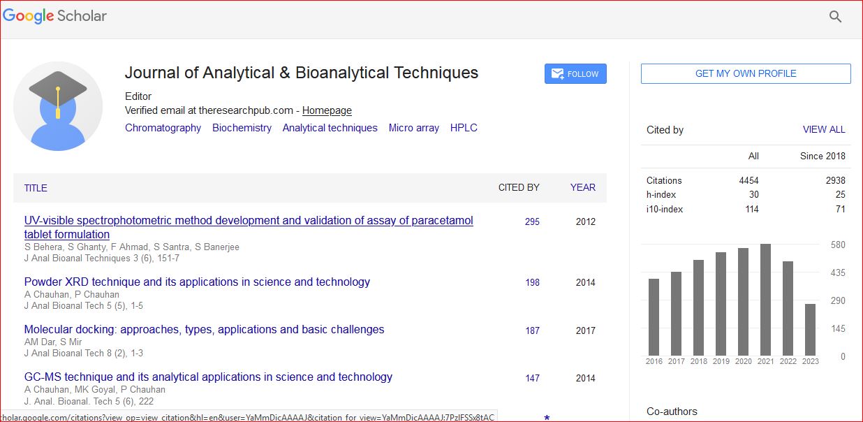
Google Scholar citation report
Citations : 6413
Journal of Analytical & Bioanalytical Techniques peer review process verified at publons

Indexed In
- CAS Source Index (CASSI)
- Index Copernicus
- Google Scholar
- Sherpa Romeo
- Academic Journals Database
- Open J Gate
- Genamics JournalSeek
- JournalTOCs
- ResearchBible
- China National Knowledge Infrastructure (CNKI)
- Ulrich's Periodicals Directory
- Electronic Journals Library
- RefSeek
- Directory of Research Journal Indexing (DRJI)
- Hamdard University
- EBSCO A-Z
- OCLC- WorldCat
- Scholarsteer
- SWB online catalog
- Virtual Library of Biology (vifabio)
- Publons
- Euro Pub
- ICMJE
Useful Links
Related Subjects
Share This Page
Nanoparticles for Magnetic Resonance Imaging
3rd International Conference and Exhibition on Analytical & Bioanalytical Techniques
Ramesh S. Chaughule
ScientificTracks Abstracts: J Anal Bioanal Techniques
Abstract
Magnetic resonance imaging (MRI) is one of the most powerful tools for non invasive clinical diagnosis due to contrast in soft tissues. It may be useful for the early detection of lesions. Protons (hydrogen nuclei) from different tissues yield different relaxation times providing a picture of anatomical pictures. Contrast agents have made a significant impact in the use of MRI for various clinical indications and improve quality of images. It is shown that inclusion of contrast agents improves the quality of images and accentuates differences between normal and diseased lesions. Since the introduction of the first MRI contrast agent Gd-DTPA in 1988, there has been a tremendous increase in the number of contrast-enhanced examinations. Nanoparticles that possess magnetic properties can be manipulated by an external magnetic field gradient and thus useful for novel biomedical applications, such as magnetic drug targeting, hyperthermia, MRI contrast enhancement and magnetic separation. Advances in nanoparticle contrast agents for molecular imaging have made magnetic resonance imaging a promising modality for noninvasive visualization and assessment of vascular and cardiac disease processes. MRI contrast agents contain paramagnetic or superparamagnetic metal ions that attract considerable interest due to their excellent properties such as large surface area and contrasting effects. The contrast agents are used primarily to increase the sensitivity of MRI for detecting various pathological processes and also for characterizing various pathologies. Since iron oxide nanoparticles have markedly higher value of the magnetic moment and thus, present much higher relaxivities than Gd-chelates. MRI contrast in soft tissues is due to differences in the proton density, spin lattice relaxation time T1 and spin-spin relaxation time T2 of the protons. The modification in contrast is due to their effect of shortening the relaxation time T1 and/or T2 of the protons located in their vicinity. If the contrast agent reduces time T1 (paramagnetic contrast agents, such as Gadolinium chelates), we observe enhancement in T1 weighted sequences. On the other hand, if it shortens T2 (superparamagnetic contrast agents such as SPIO and USPIO), there will be a reduction in the T2 and T2* signal. Under an applied magnetic field, induced magnetic spins in magnetic nanoparticles perturb the nuclear spin relaxation processes of protons of water molecules surrounding magnetic nanoparticles. This effect leads to the shortening of spin-spin relaxation time (T2) of the protons due to inhomogeneities in local magnetic field and fluctuating magnetic fields at molecular level, which results in darkening of MR images. Thus regions containing SPIO contrast agents appear darker in an MRI than regions without the agent. For example, when SPIOs are delivered to the liver, unlike diseased cells, healthy liver cells can uptake the particles and darkened. The basis of T1 weighted imaging is the longitudinal relaxation. A T1 weighted magnetic resonance image is created typically by using short TE and TR times. Due to the larger longitudinal and transverse magnetization, fat has a higher signal and will appear bright on a T1 contrast MR image. Conversely, water has less longitudinal magnetization prior to a RF pulse, therefore less transverse magnetization after a RF pulse yielding low signal appearing dark on a T1 contrast image. A brief review of superparamagnetic iron oxide (SPIO) based MRI contrast agents and their current clinical applications are presented. The effect of relaxation times of magnetic nanoparticles with varying size, surface modification etc on the development of contrast in MRI will also be discussed.Biography
Ramesh Chaughule was senior Scientist at the Tata Institute of Fundamental Research, Mumbai and now is Adjunct Professor at Ramnarain Ruia College, Mumbai. Also he is Research Advisor at Gogate-Jogalekar College, Ratnagiri, Maharashtra. He has served on International Atomic Energy Agency, Vienna and obtained several assignments out side India. He has more than 60 publications in reputed international journals. In addition he is chief Editor of 3 books in the field of Nanotechnology published by American Scientific Publishers, USA and one book on MRI.