Constrictive Pericarditis with Extensive Pericardial Calcification: Case Report and Review of Literature
Received: 05-Oct-2021 / Accepted Date: 19-Oct-2021 / Published Date: 26-Oct-2021 DOI: 10.4172/jcpr.1000147
Abstract
Constrictive pericarditis refers to inflammation of the pericardial sac, possibly leading to acute heart failure. More than 80% cases are presumed to be due to recent or remote viral illnesses. Prominent features include chest pain, dyspnea and Electrocardiogram (ECG) revealing P-R segment depression, diffuse concave ST segment elevation, and T-wave inversion. Echocardiogram and cardiac Magnetic Resonance Imaging (MRI) can help establish diagnosis. Over time, the pericardium can undergo fibrosis or calcification resulting in excessive symptoms. After medical management with ibuprofen, colchicine or steroids, partial or complete pericardiectomy is considered. We are presenting a case with constrictive pericarditis due to extensive pericardial calcification, and ultimate resolution with pericardiectomy.
Keywords: Constrictive pericarditis; Coxsackievirus; Pericardial calcification; Pericardiectomy
Introduction
Constrictive pericarditis refers to inflammation of the pericardial sac, possibly leading to acute heart failure. More than 80% cases are presumed to be due to recent or remote viral illnesses. Prominent features include chest pain, dyspnea and ECG revealing P-R segment depression, diffuse concave ST segment elevation, and T-wave inversion. TTE and cardiac MRI can help establish diagnosis. Over time, the pericardium can undergo fibrosis or calcification resulting in excessive symptoms. After medical management with ibuprofen, colchicine or steroids, partial or complete pericardiectomy is considered. We are presenting a case with constrictive pericarditis due to extensive pericardial calcification, and ultimate resolution with pericardiectomy.
Case Report
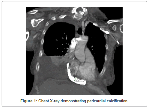
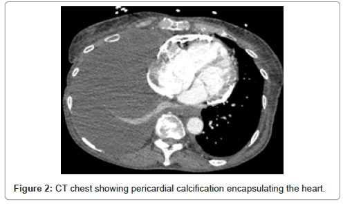
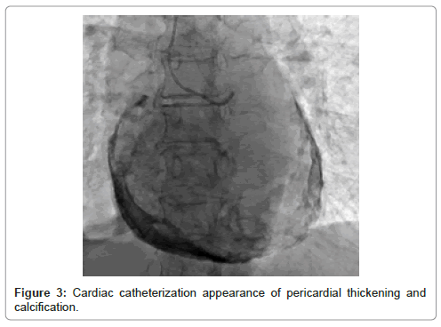
A 68-year-old female with right heart failure leading to non- alcoholic liver cirrhosis and paroxysmal atrial fibrillation presented to the emergency department with worsening dyspnea, weight gain, exertional chest tightness and increased fatigue for one week. Due to insurance challenges, she was non-adherent to her medications. Physical examination revealed temperature 98 F, respiratory rate 24 breaths/minute, pulse 114 beats/minute, blood pressure 154/92 mm Hg, jugular venous distension, distended abdomen and 3+pitting edema in both legs. On laboratory examination, hemoglobin, white blood cell count and platelet count were normal. Troponin was normal, pro-BNP was 1268 ng/mL and ECG showed atrial fibrillation with rapid ventricular response. Chest X-ray and computed tomography of the chest and abdomen revealed dense pericardial calcification (Figures 1 and 2), large right-sided pleural effusion, liver cirrhosis and anasarca. Transthoracic echocardiogram showed a new decreased left ventricular ejection fraction of 40-45%, flattened interventricular septum and severely enlarged right atrium. Cardiac catheterization showed normal epicardial coronary arteries, myocardial bridging in mid-Left Anterior Descending (LAD) segment, substantial pericardial calcification and discordance between simultaneous left and right ventricular pressure (Figure 3). It was discovered that the patient had a history of coxsackievirus infection in childhood. Constrictive Pericarditis (CP) secondary to prior coxsackievirus infection was diagnosed. Patient was treated with guideline-directed medical therapy with metoprolol, atorvastatin for heart failure and showed clinical improvement at discharge. She was then admitted later for worsening shortness of breath and was treated with digoxin for atrial fibrillation, and intravenous furosemide. Paracentesis was performed with removal of 850-mL clear transudative fluid. Patient had undergone paracentesis with removal of 4350-mL clear transudative fluid 1 month earlier. Pericardiectomy was planned and intra-operatively, the patient was noted to have a greater than 2 cm thick and calcified pericardium that completely encased the heart. The posterior pericardium was more than 4 cm. Mechanical cardiac bypass was used briefly and complete pericardiectomy was performed. A 32-French chest tube was placed in the anterior mediastinum, 28-French chest tubes were placed in the right and left pleural spaces, and a 10-French JP was placed in the posterior pericardium. Ventricular wires were placed, secured to skin and connected to the pacemaker device. The patient was intubated during the procedure, and moved to the intensive care unit after the procedure. She was extubated after 2 days. Chest tubes were removed POD 15-17. The temporary pacemaker wires were clipped on discharge. One month later, her symptoms resolved, and echocardiography showed LVEF 60-65%.
Discussion
CP refers to the process of inflammatory and fibrotic loss of elasticity and compliance of the pericardium, a sack of mesothelial cells and collagen that envelope the heart, resulting in diminished diastolic filling, causing acute diastolic heart failure [1,2]. A report in 1962 noted that approximately 50% of cases of CP were caused by Tuberculosis in North America. Worldwide, the causes included Human Immunodeficiency Virus (HIV) and AIDS [2]. More recent studies of the etiologies of CP have revealed other causes. In a retrospective study of 57 patients, prior cardiothoracic surgery was found to be the most common cause [3]. 80- 90% of idiopathic cases are presumed to be due to viral etiologies [4]. Common viruses include enteroviruses, including coxsackievirus and echovirus, herpesviruses, adenoviruses and parvovirus B19 [4]. While the pathogenesis of viral CP is not completely understood, evidence suggests a role of auto-antibodies against cells of the pericardium, including anti-heart antibodies and anti-intercalated disk antibodies [2].
Symptoms of pericarditis can frequently be confused with other cardiac pathologies. Pericarditis has been noted in 5% of patients presenting to the emergency department with chest pain [5]. Classically, chest pain in pericarditis has been noted to be postural, and improving with leaning forward. A pericardial rub may also be auscultated, and there is an additional risk for the development of a pericardial effusion. Pericarditis may also be associated with specific ECG findings, including P-R segment depression, diffuse concave ST segment elevation, and T-wave inversion [5]. Echocardiography is the mainstay imaging modality for pericarditis; however, Gadolinium-Enhanced (GE) Cardiac Magnetic Resonance Imaging (CMR) is also used. Typical MRI findings show late GE of the pericardium with absence of edema and GE of the myocardial tissue [2,5]. Troponin and Brain Natriuretic Peptide (BNP) are not typically noted to be elevated [5].
Echocardiographic findings for cases of acute CP are normal in 40% of cases of acute pericarditis [5], but help rule out cardiac tamponade and determine the degree of constriction and severity of diastolic dysfunction. In many patients, including ours, CP may precipitate a pericardial effusion, further exacerbating pericardial inflammation and heart failure. Recently, advances in 3-dimensional echocardiography have also allowed the rapid and reliable determination of pericardial effusion location. Echocardiography is also utilized for visualization of the pericardium for instances requiring a pericardiocentesis in effusive cases of pericarditis [5].
Management of CP involves managing the inflammatory processes affecting the pericardium, and relieving the pressure generated on the heart from the pericardium. To reduce inflammation, NSAIDS have shown clinical benefit, though no Randomized Controlled Trial (RCT) has validated their efficacy for CP cases. RCTs, notably, have also shown that administration of colchicine helps reduce symptoms at 72 hours after administration (COPE trial) and reduce the rates of recurrence (CAFE-IP trial) [5]. For healthy patients, mild constriction in CP can often be managed effectively with diuretics; however, for most patients with CP, pericardiectomy is a definitive treatment option [6]. Pericardiectomy involves the surgical resection of the pericardial sac enveloping the heart. While complete pericardiectomy has shown to be the most optimal in improving mortality in patients with CP, subtotal resection has also shown definitive improvement in symptomology and mortality outcomes [6]. In a retrospective study of pericardiectomy [6], 79% of CP patients receiving surgery had New York Heart Association class I or II for heart failure, showing satisfactory outcomes. In-hospital mortality was at 9%, while overall survival rate was 87% at 5 years and 78% at 10 years.
Conclusion
CP is an entity leading to features of pericarditis and acute onset of heart failure. Currently, viral etiologies with prior viral illness are considered major causes. Transthoracic echocardiogram and cardiac MRI help in establishing the diagnosis. Effective surgical management includes radical pericardiectomy. An important inter-departmental communication and involvement between cardiology and cardiothoracic surgeons is essential for prompt and complete treatment.
Funding
Not applicable.
References
- Jaworska-Wilczynska M, Trzaskoma P, Szczepankiewicz AA, Hryniewiecki T (2016) Pericardium: structure and function in health and disease. Folia Histochem Cytobiol 54: 121-125.
- Miranda WR, Oh JK (2017) Constrictive pericarditis: a practical clinical approach. Prog Cardiovasc Dis 59: 369-379.
- Landex NL, Ihlemann N, Olsen PS, Gustafsson F (2015) Constrictive pericarditis in a contemporary danish cohort: aetiology and outcome. Scand Cardiovasc J 49: 101-108.
- Imazio M, Gaita F, LeWinter M (2015) Evaluation and treatment of pericarditis: a systematic review. Jama 314: 1498-1506.
- Chiabrando JG, Bonaventura A, Vecchié A, Wohlford GF, Mauro AG, et al. (2020) Management of acute and recurrent pericarditis: JACC state-of-the-art review. J Am Coll Cardiol 75: 76-92.
- Vistarini N, Chen C, Mazine A, Bouchard D, Hebert Y, et al. (2015) Pericardiectomy for constrictive pericarditis: 20 years of experience at the Montreal Heart Institute. Ann Thorac Surg 100: 107-113.
Citation: Mahajan P, Naik A, Aedma SK, Mehta R, Ally S, et al. (2021) Constrictive Pericarditis with Extensive Pericardial Calcification: Case Report and Review of Literature. J Card Pulm Rehabil 5: 147. DOI: 10.4172/jcpr.1000147
Copyright: © 2021 Mahajan P, et al. This is an open-access article distributed under the terms of the Creative Commons Attribution License, which permits unrestricted use, distribution, and reproduction in any medium, provided the original author and source are credited.
Select your language of interest to view the total content in your interested language
Share This Article
Open Access Journals
Article Tools
Article Usage
- Total views: 3328
- [From(publication date): 0-2021 - Dec 19, 2025]
- Breakdown by view type
- HTML page views: 2562
- PDF downloads: 766


