Research Article Open Access
Observation of the Pharynx to the Cervical Esophagus Using Trans-nasal Endoscopy with Image-enhanced Endoscopy for Early Detection of Head and Neck Cancers
Kenro Kawada1*, Takuya Okada1, Taro Sugimoto2, Kazuya Yamaguchi1, Yuudai Kawamura1, Toshihiro Matsui1, Masafumi Okuda1, Taichi Ogo1, Yuuichiro Kume1, Andres Mora1, Akihiro Hoshino1, Yutaka Tokairin1, Yasuaki Nakajima1, Ryuhei Okada2, Yusuke Kiyokawa2, Fuminori Nomura2, Yosuke Ariizumi2, Takahiro Asakage2, Takashi Ito3 and Tatsuyuki Kawano41Department of Gastrointestinal Surgery, Tokyo Medical and Dental University, Tokyo, Japan
2Department of Head and Neck surgery, Tokyo Medical and Dental University, Tokyo, Japan
3Department of Human Pathology, Tokyo Medical and Dental University, Tokyo, Japan
4Department of Surgery, Soka Municipal hospital, Saitama, Japan
- Corresponding Author:
- Kawada K
Department of Gastrointestinal Surgery
Tokyo Medical and Dental University, Tokyo, Japan
Tel: +81-3-5803-5254
Fax: +81-3-3817-4126
E-mail: kawada.srg1@tmd.ac.jp
Received Date: August 18, 2017; Accepted Date: August 29, 2017; Published Date: August 31, 2017
Citation: Kawada K, Okada T, Sugimoto T, Yamaguchi K, Kawamura Y, et al. (2017) Observation of the Pharynx to the Cervical Esophagus Using Trans-nasal Endoscopy with Image-enhanced Endoscopy for Early Detection of Head and Neck Cancers. J Gastrointest Dig Syst 7:522. doi:10.4172/2161-069X.1000522
Copyright: © 2017 Kawada K, et al. This is an open-access article distributed under the terms of the Creative Commons Attribution License, which permits unrestricted use, distribution, and reproduction in any medium, provided the original author and source are credited.
Visit for more related articles at Journal of Gastrointestinal & Digestive System
Abstract
Introduction: We started endoscopic treatment for superficial pharyngeal cancer in 1996, and thus far, 97 lesions of 77 cases of superficial head and neck cancer have been detected using trans-oral endoscopy. However, some areas are difficult to observe with trans-oral endoscopy because of the gag reflex. We have therefore applied transnasal endoscopy for observing of the pharynx to the cervical esophagus.
Methods: To avoid overlooking cancers located at the floor of the mouth, soft palate and uvula, we first observe the oral cavity. After administering local anesthesia to the nose without sedation, the endoscope is inserted through the nose. When the tip of the endoscope reaches caudal to the uvula, the patient opens his or her mouth wide, sticks the tongue forward as far as possible and makes a makes a vocalization like “ayyy”. The endoscopist then makes the endoscope take a U-turn and observes the oropharynx, particularly radix linguae. To examine the hypopharynx and the orifice of the esophagus, the patient is asked to blow hard and puff their cheeks while the mouth remains closed. This approach provides a much better view of the orifice of the esophagus than is possible with trans-oral endoscopy with deep sedation.
Results: In this study, we detected 22 superficial cancers of the oral cavity. Previous efforts to detect such cancers using trans-oral endoscopy have failed. In addition, we were never able to detect early cancers located at base of tongue in the past, but since implementing the intra-oropharyngeal U-turn method, we have detected more than 10 cases. We were also never able to detect early cancers located at the pharyngoesophageal junction in the past, but since implementing the modified Valsalva maneuver, we have detected more than 20 cases. Between 2008 and 2016, a total of 164 cases of 227 lesions of superficial head and neck cancer were detected by trans-nasal endoscopy, which is more than twice as many as were detected with conventional screening. Mucosal redness, white deposits or loss of a normal vascular pattern and proliferation of vascular pattern such as small dots or salmon roe with a close-up view of it are important characteristics to diagnose superficial pharyngeal cancer. Moreover, a brownish area using image-enhanced endoscopy is useful for early diagnosis. With adequate extension of the pharyngeal mucosa using the Valsalva maneuver, observing the protruded areas should prove useful for diagnosing the depth of invasion.
Conclusions: Observing the pharynx to the cervical esophagus using trans-nasal endoscopy with imageenhanced endoscopy is useful for early detection of head and neck cancers.
Keywords
Trans-nasal endoscopy; Narrow band imaging; Blue laser imaging; Superficial head and neck cancer
Introduction
Field cancerization concept is described for fisrt time in 1953, old concept and still available [1]. According to the concept, head and neck cancers, especiallypharyngeal cancer, frequently coexist with esophageal cancer [2]. Patients selected as high-risk group in pharyngeal cancer, have important benefits from endoscopic suveiliance.
We started endoscopic treatment for superficial pharyngeal cancer in 1996, and thus far, 97 lesions of 77 cases of superficial head and neck cancer have been detected using trans-oral endoscopy [3]. However, some areas are difficult to observe with trans-oral endoscopy because of the gag reflex. We have therefore applied new techniques for observing of the pharynx to the cervical esophagus [4].
Trans-nasal endoscopy hasrapidly become widespread due to lower degree of discomfort and the lesser impact on cardio-pulmonary system. However, it has been a problem that in image quality and maneuver ability ultrathin endoscopy is inferior to trans-oral endoscopy. But recently ultrathin endoscopes have made tremendous progress in the past ten years. The latest model has high resolution that is equal to that of a conventional endoscope. Our aim is to introduce the endoscopic screening methods, in patients with the coexistence of superficial head and neck cancers and esophageal cancer in Japan.
Methods
We applied trans-nasal endoscopic approach for observing of pharynx to the cervical esophagus in 2008. The modified Valsalva maneuver and oral cavity screening started in 2009the intraoropharyngeal U-turn method started in 2012. The development of trans-nasal endoscopy with image enhanced endoscopyNarrow Band Imaging (NBI) [5-8]. Blue Laser Imaging (BLI) and Linked Color Imaging (LCI), now enables the wider observation and can be used to obtain adequate information for diagnosing early pharyngeal cancers without magnification [9,10].
Equipment
The upper endoscopes used were GIF-290N and GIF-260NS (Olympus, Co., Tokyo, Japan) or EG-530NW, EG-580NW, EGL580NW, (Fujifilm, Co., Tokyo, Japan). Ultrathin endoscopy using EG-580NW, EG-L580NW allows detailed observation in near views because these endoscopes have 3 mm of focal length to achieve good endoscopic images.
These scopes have an increased resolution of CCD, which produces structural image of superficial head and neck cancer distinct from the surrounding mucosa. Recently, image-enhanced endoscopy (IEE) has been carried out to diagnose gastrointestinal tumors. Lugol chromoendoscopy can be used to detect superficial esophageal cancer, but it cannot be used for head and neck squamous cell carcinoma screening because of the risk of aspiration. The narrow band imaging (NBI) system is an image-enhanced technology that uses NBI filters [5].
Recently several reports have indicated the possibility of narrowband imaging (NBI) endoscopy with magnification improving the detection of superficial pharyngeal cancer [6,7]. Compared with conventional endoscopy, the detection of superficial lesion is dramatically improved and the microvascular structure of the mucosal surface in significantly enhanced by NBIA rhinolaryngoscope equipped with an NBI system is also available for examining these lesions [8].
The LASERIO system (Fuji Film, Co.TokyoJapan) is the new generation gastrointestinal endoscopy system to use laser light as illumination. They use two lasers, and one white light phosphor to accomplish the visual enhancement of the surface vessels of the mucosa. One of lasers have a wavelength of 450 nm, it stimulates the phosphor to irradiate white color illumination. And the other laser uses a wavelength of 410 nm to enhance the blood vessels to show the deepness of the mucosa [9].
The linked color imaging (LCI) function is a new image enhancing technology available for the LASERIO system that makes slight differences in mucosal color tones easier to recognize [10].
Screening Technique
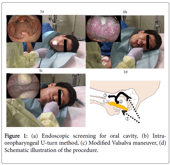
Prior to trans-nasal endoscopy, each nasal cavity is sprayed with 0.5 ml of 0.05% naphazoline nitrate for vasoconstriction, and 2 ml of 4% lignocaine solution for topical anesthesia. Our screening procedure is as follows: the patient is asked to bow their head deeply in the left lateral position, and then we keep our hand on the back of the patient’s head and push it forward by one hand-span. The patient is then asked to lift up their chin as far as possible (lateral sniffing position). To avoid overlooking cancers located at the floor of the mouth, soft palate and uvula, we first observe the oral cavity (Figure 1a). We then insert the endoscope without a mouthpiece and subsequently observe the upper, lateral and posterior wall of the oropharynx while the patient sticks their tongue out. After observing the buccal cavity, further oropharyngeal observation is carried out with a retroflexed endoscope is inserted through the nose. When the tip of the endoscope reaches caudal to the uvula, the patient opens his or her mouth wide, sticks the tongue forward as far as possible (Figure 1b) and makes a makes a vocalization like “ayyy”. The endoscopist then makes the endoscope take a U-turn (intra-oropharyngeal U-turn method and observes the oropharynx, particularly radix linguae [11]. To examine the hypopharynx and the orifice of the esophagus, the patient is asked to blow hard and puff their cheeks while the mouth remains closed modified Valsalva maneuver [12,13] (Figure 1c). The post-cricoid subsite of hypopharynx had lifted completely. This procedure provided a much better view of the orifice of the esophagus than had been possible with trans-oral endoscopy. Figure 1d is a schema of the procedure. Table 1 is a summary of the screening procedure.
| Screening procedure |
| Lateral sniffing position, no sedation |
| Observing oral cavity without mouth piece |
| Intra-oropharyngeal U-turn method (stick the tongue forward) |
| Modified Valsalvamaneuver (blow hard and puff the cheeks) |
Table 1: Summary of the screening procedure.
Results
According to the General rules for clinical studies on head and neck cancer in Japan, carcinoma in situ and subepithelial cancers with or without lymph node metastasis, are defined as a superficial cancer [14].
In this study, we detected 22 superficial cancers of the oral cavity. Previous efforts to detect such cancers using trans-oral endoscopy have failed. In addition, we were never able to detect early cancers located at base of tongue in the past, but since implementing the intraoropharyngeal U-turn method, we have detected more than 10 cases [15].
We were also never able to detect early cancers located at the pharyngoesophageal junction in the past, but since implementing the modified Valsalva maneuver, we have detected more than 20 cases. Between 2008 and 2016, a total of 164 cases of 227 lesions of superficial head and neck cancer were detected by trans-nasal endoscopy, which is more than twice as many as were detected with conventional screening (Table 2).
| Transoral (77) 1996-2016 | Transnasal (164) 2008-2016 | |
|---|---|---|
| Cases | Male: 72 | Male: 156 |
| Female: 5 | Female: 8 | |
| lesions | 97 | 227 |
| Hypopharynx | 66 | 130 |
| Piriform sinus | 41 (rt19-lt22) | 80 (rt42-lt38) |
| Posterior wall | 24 | 39 |
| Postcricoid | 1 | 11 |
| Oropharynx | 24 | 58 |
| lateral wall | 2 | 12 |
| anteriorwall | 2 | 14 |
| posterior wall | 15 | 22 |
| Superior wall | 5 | 10 |
| Larynx | 6 | 17 |
| Oral cavity | 1 | 22 |
| Floor of mouth | 0 | 11 |
| Tongue | 1 | 7 |
| Buccal mucosa | 0 | 4 |
Table 2: The superficial head and neck cancers detected by trans-oral endoscopy and trans-nasal endoscopy.
Mucosal redness, white deposits or loss of a normal vascular pattern and proliferation of vascular pattern such as small dots or salmon roe with a close-up view of it are important characteristics to diagnose superficial pharyngeal cancer.
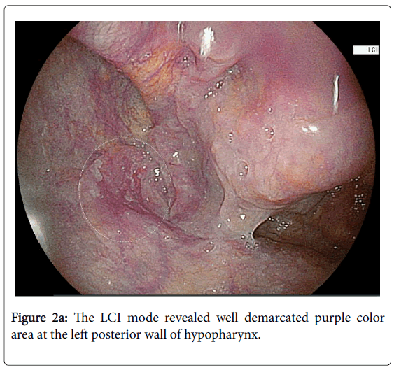
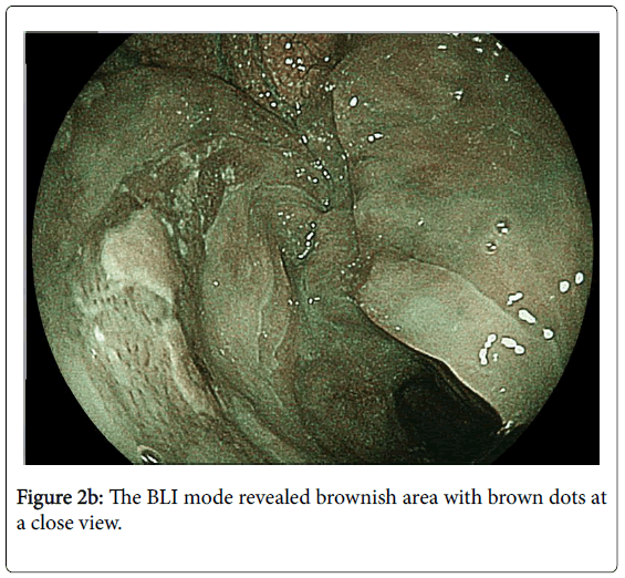
The best method for detecting a lesion is to slowly move the scope, and pay special attention to slight differences in the well demarcated area. LCI is useful to get a distant viewand BLI is useful to get a close-up view (Figures 2a and 2b).
Case Presentation
Case 1
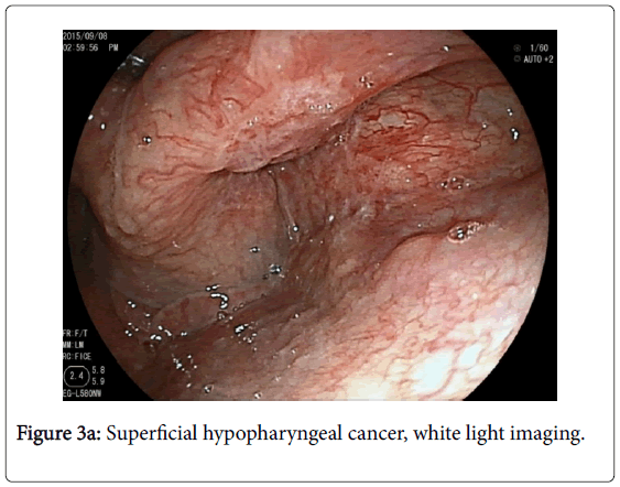
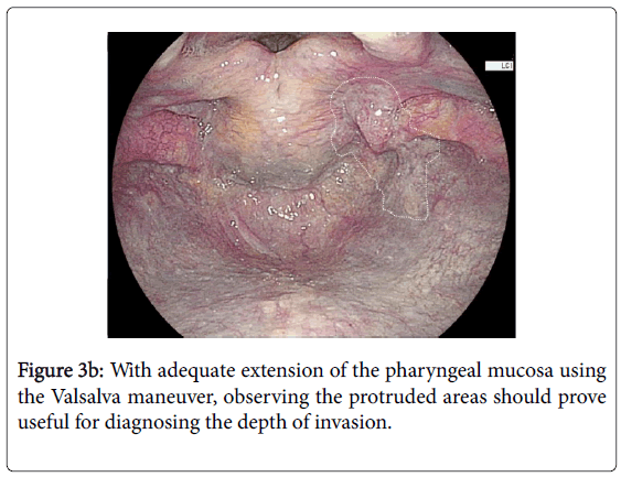
There was a white irregular mucosa with loss of a normal vascular pattern at the right piriform sinus (Figure 3a). Using modified Valsalva maneuver, the full extension of pharyngeal mucosa was obtained that the clear view of the tumor was seen. With adequate extension of the pharyngeal mucosa using the modified Valsalva maneuver, observing the protruded areas should prove useful for diagnosing the depth of invasion (Figure 3b).
Case 2
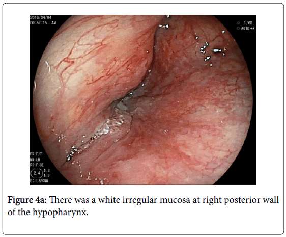
There was a white irregular mucosa at right posterior wall of the hypopharynx (Figure 4a). We could not observe the whole view. Using modified Valsalva maneuver, we could diagnose that the tumor was located at the center of posterior wall of the hypopharynx. The distal part of the tumor closed to the cervical esophagus (Figure 4b).
Discussion
According to the “field cancerization” concept, head and neck cancer, especially pharyngeal cancer, frequently coexist with esophageal cancer [1]. It has been reported that the application of magnifying endoscopy using the NBI system changes the diagnostic strategy for the early detection of early pharyngeal cancers [6]. NBI works by exploiting the wavelength selectivity of the absorption characteristics of hemoglobin. The superiority of NBI was also recently demonstrated in a multicenter randomized controlled trial in Japan [7]. Howeversome areas are difficult to observe with trans-oral endoscopy. In particular, achieving circumferential observation of the hypopharyngeal mucosa is difficult during conventional endoscopy due to the anatomically closed field, effects of the gag reflex and accumulation of saliva.
On conventional endoscopic screening, the physician usually inserts the endoscope from the left pyriform sinus of the hypopharynx to the cervical esophagus,with a blind space in the posterior wall and postcricoid subsite of the hypopharynx as well as radix linguae. Therefore, detecting early signs of cancer in the blind space is difficult for gastrointestinal endoscopists. On the other hand, trans-nasal endoscopy may be performed comfortably due to attenuation of the gag reflex. In Japan, the transnasal endoscopy is a very popular procedure and can be performed without sedation. Since we developed the pharyngo-laryngeal observation method using trans-nasal endoscopy in 2009, we have constantly evolved the procedure in order to better detect carcinoma in the head and neck at earlier stages in cases often coexistent with esophageal cancer [4]. In 2014, a new transnasal endoscopy device with Blue laser Imaging (BLI) was developed. Furthermore, Linked Color Imaging (LCI) was developed. The LCI mode allows for detailed observation from a distance. The new generation image-enhanced endoscopy is also useful as well as NBI system.
In this retrospective study, between 2008 and 2016, a total of 164 cases of 227 lesions of superficial head and neck cancer were detected by trans-nasal endoscopy, which is more than twice as many as were detected with conventional screening. This study is not prospective, not randomized, and performed by one institution, so there is a limitation. But, the method for wide visualization of pharynx provided an excellent endoscopic field of pharynx which is not possible using conventional method. If early stage cancers can be detected with this screening method, the prognosis of the patients will be improved. While this technique has not yet caught on in Japan, it is very easy to perform and not expensive, so we recommend screening the high-risk patients, which include heavy drinkers, heavy smokers, and those with esophageal cancer.
Conclusions
Observation of the pharynx to the cervical esophagus using transnasal endoscopy with image-enhanced endoscopy is useful for early detection of head and neck cancers.
References
- Slaughter DP, Southwick HW, Smejkal W (1953) Field cancerizationin oral stratified squamous epithelium: Clinical implications of multicentric origin. Cancer 6: 963-968.
- McGuirt WF, Matthews B, Koufman JA (1982) Multiple simultaneous tumors in patients with head and neck cancer. Cancer 50: 1195-1199.
- Nagai K, Kawada K, Nishikage T (2003) Endoscopic treatment for hypopharyngeal cancer.Stomach and Intestine 38: 331-338.
- Kawada K, Kawano T, Sugimoto T (2013) Key points and techniques for trans-nasal endoscopic screening for superficial hypopharyngeal cancer. Treatment Strategies Gastroentel 2:42.
- Gono K, Yamazaki K, Doguchi N (2003) Endoscopic observation of tissue by narrow band illumination. Opt Rev10: 1-5.
- Muto M, Nakane M, Katada C (2004) Squamous cell carcinoma in situ in oropharyngeal and hypopharyngeal mucosal sites. Cancer 101: 1375-1381.
- Muto M, Minashi K, Yano T (2010) Early detection of superficial squamous cell carcinoma in the head and neck region and esophagus by narrow band imaging: A multicenter randomized controlled trial. JClinOncol 28: 1566-1572.
- Watanabe A, Tsujie H, Taniguchi M (2006) Laryngoscopic detection of pharyngeal carcinoma in situ with narrow band imaging. Laryngoscope 116: 650-654.
- Kawada K, Kawano T, Sugimoto T (2015) Observation of the pharynx to the cervical esophagus using transnasal endoscopy with blue laser imaging. SomchaiAmornyotin(eds), Endoscopy-Innovative Uses and Emerging Technologies. 1st(ed)Intech, Croatia pp: 215-231.
- DohiO, Yagi N, Onozawa Y(2016) Linked color imaging improves endoscopic diagnosis of active Helicobacter pylori infection. EndoscInt Open E800-E805.
- Kawada K, Okada T, Sugimoto T (2014) Intraoropharyngeal U-turn method using transnasal esophagogastroduodenoscopy. Endoscopy 46: E137-138.
- Spraggs PD, Harris ML (1995) The modified Valsalvamaneuver to improve visualization of the hypopharynx during flexible nasopharyngoscopy. J LaryngolOtol109: 863-864
- Kawada K, Kawano T,Sugimoto T (2015) Case of a superficial hypopharyngeal cancer at the pharyngoesophageal junction which is detected by transnasal endoscopy using trumpet maneuver.Open J of Gastroenterol5: 1-6.
- General Rules for Clinical Studies on Head and Neck Cancer. Japan Society for Head and Neck Cancer. 5th(ed), Kanehana, Japanpp: 38-45.
- Kawada K, Kawano T, Okada T (2017) The usefulness of an Intra-oropharyngealu-turnmethod using trans-nasal endoscopy for detecting superficial squamous cell carcinoma of the base of the tongue. J Otolaryngol ENT Res8:00240.
Relevant Topics
- Constipation
- Digestive Enzymes
- Endoscopy
- Epigastric Pain
- Gall Bladder
- Gastric Cancer
- Gastrointestinal Bleeding
- Gastrointestinal Hormones
- Gastrointestinal Infections
- Gastrointestinal Inflammation
- Gastrointestinal Pathology
- Gastrointestinal Pharmacology
- Gastrointestinal Radiology
- Gastrointestinal Surgery
- Gastrointestinal Tuberculosis
- GIST Sarcoma
- Intestinal Blockage
- Pancreas
- Salivary Glands
- Stomach Bloating
- Stomach Cramps
- Stomach Disorders
- Stomach Ulcer
Recommended Journals
Article Tools
Article Usage
- Total views: 5519
- [From(publication date):
August-2017 - Aug 23, 2025] - Breakdown by view type
- HTML page views : 4560
- PDF downloads : 959

