Research Article Open Access
Occurrence Regions and Sites of Peri-implant Inflammation With Bone Resorption in Partially-edentulous Patients
Motohiro Munakata*, Noriko Tachikawa, Katsuichiro Maruo, Aoi Sakuyama, Yoko Yamaguchi and Shohei Kasugai
Associate Professor, Oral Implantology, Yokosuka, Kanagawa Japan
- *Corresponding Author:
- Motohiro Munakata
Associate Professor
Oral Implantology, Yokosuka
Kanagawa Japan
Tel: 81 46 8228880
E-mail: munakata@kdu.ac.jp
Received Date: June 25, 2014; Accepted Date: July 25, 2014; Published Date: August 02, 2014
Citation: Munakata M, Tachikawa N, Maruo K, Sakuyama A, Yamaguchi Y, et al. (2014) Occurrence Regions and Sites of Peri-implant Inflammation With Bone Resorption in Partially-edentulous Patients. J Oral Hyg Health 2:146. doi:10.4172/2332-0702.1000146
Copyright: © 2014 Munakata M, et al. This is an open-access article distributed under the terms of the Creative Commons Attribution License, which permits unrestricted use, distribution, and reproduction in any medium, provided the original author and source are credited.
Visit for more related articles at Journal of Oral Hygiene & Health
Abstract
Aim: The aim of this study was to clarify the occurrence regions and sites of peri-implant bone resorption and inflammation in partially-edentulous patients.
Methods: Five hundred one partially-edentulous patients with 738 implants in function for more than 5 years were included in this study for the evaluation of the bone resorption by using dental radiograph and probing. Considering physiological bone remodeling, the mean mesio-distal bone resorption around the implant was measured on dental radiograph.
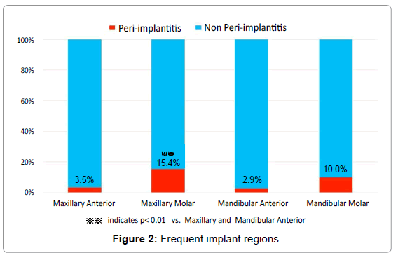
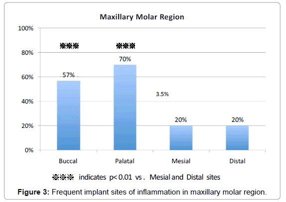
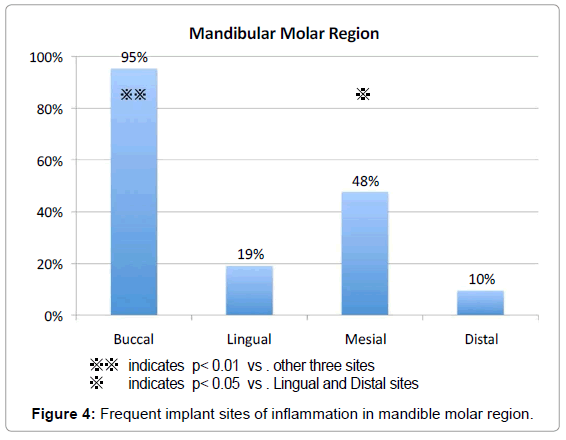
Results: In 65 patients (13.0% of the total patients) with 76 implants (10.3% of the total implants), peri-implant bone resorption was identified. The mean functional loading time of these implants was 8.4 years. Occurrence regions were frequently found in the molar regions in maxilla (15.4%) and the molar region in mandible (10.0%) In these leisions detected radiologically, the bleeding on probing was seen in 95.2% of the buccal sites in mandibular molar regions, 70.0% of the palatal sites in maxillary molar regions and 56.7% of the buccal sites in maxillary molar regions with statistically significant differences.
Conclusion: From the limitation of the information in this study, it was concluded that the sites that tend to be vulnerable to peri-implant inflammation were the buccal site in mandible, and the buccal and palatal sites in maxilla.
Keywords
Bone resorption; Dental implant; Dentistry; Peri-implant ; Peri-implantitis
Introduction
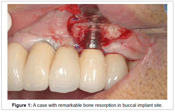
Dental implants have been successfully used in the treatment of complete and partial edentulous patient subjects [1]. Nevertheless, dental implant failures have also been reported [2,3]. These failures are classified on the basis of chronology, i.e. early or late failure. Early dental implant failures are attributed to surgical trauma, inadequate bone volume, lack of primary stability, intra-osseous infection, and bacterial contamination of the recipient site [3,4]. Late dental implant failures are associated with peri-implantitis and/or biomechanical overload [2,3,5]. In the Sixth European Workshop on Periodontology, peri-implant disease was a collective term for inflammatory reactions in the tissues surrounding an implant [6,7]. “Peri-implant mucositis” is defined as inflammation of the mucosa around an implant without loss of supporting bone, while “peri-implantitis” is characterized by loss of supporting bone together with mucosa inflammation. It has been reported that peri-implant mucositis occurs in 80% of the subjects and in 50% of the implant sites and that peri-implantitis is identified in 28% and 56% of subjects and in 12% and 43% of implant sites, respectively [7]. As potential risk factors for peri-implantitis, Heitz-Mayfield [8] listed the history of periodontal disease, diabetes mellitus, smoking, oral hygiene condition, alcohol intake, genotype, presence of cornified mucosa, and the implant surface property. Oral hygiene condition, history of periodontal disease, smoking, and diabetes mellitus, etc., have been reported as related risk factors. Thus, the disease will be obviously more frequent in the future, as long as a specific therapy or prevention will not establish. Clinically, bleeding and/or suppuration following probing has been proposed as a valuable clinical sign for the diagnosis of both peri-implant mucositis and peri-implantitis, while the concomitant detection of marginal peri-implant bone loss in radiographs will distinguish periimplantitis from mucositis [7]. Radiographic techniques including panoramic tomography and intra-oral radiography with long cone paralleling techniques have been widely used to monitor marginal bone levels around implants and diagnose interproximal bone loss [9]. However, conventional radiography does not enable to monitor facial and lingual/palatal bone levels around the implants being insensitive in detecting early bone changes and underestimating bone loss [10,11]. In clinical situations, cases where suppuration is found only on the buccal side or lingual/palatal sites, cases with bleeding on probing , or cases with advanced bone resorption on the buccal and lingual/palatal sites are often experienced (Figure 1). The aim of this study was to clarify the occurrence regions and sites of peri-implant bone resorption and inflammation in partially-edentulous patients.
Materials and Methods
The present clinical study was approved by the Ethical Committee, Faculty of Dentistry, Tokyo Medical and Dental University, and the written informed consents were obtained from all the patients. Subjects were 501 partial edentulous patients (738 implants) who received superstructure more than 5 years ago. All the patients who had implants inserted and superstructures made at Dental Hospital, Tokyo Medical and Dental University, between 1999 and 2006, were examined. Severe illness, uncontrolled diabetes, untreated periodontal disease and a history of head and neck radiation were excluded from the analysis. Probing pocket depth (PPD) and bleeding on probing (BOP) in the peri-implant sulcus where bone resorption was observed on the dental radiograph were explored with the 4-point method. In addition, the mean height of vertical bone defects at the both sites of the mesial and distal areas of implants was measured from the dental radiographic evaluation at least 1 year after the placement of the superstructures, since the reference time point should be considered of the bone remodeling after loading. In the radiographs the distance between the reference point and the most coronal position of bone to implant contact was assessed at the both of mesial and distal aspects of the 76 implants using a magnifying lens (10X) with a 0.1 mm graded scale. Peri-implantitis was diagnosed when the bone resorption was pictured as 2 mm or larger on dental radiograph and further BOP was observed in the peri-implant sulcus.
Data analyses
Reference of peri-implantitis in different four regions.
Examination of BOP in implant sites
Man-Whitney U-test was conducted for comparisons between different regions and sites. A p-value less than 0.05 were considered statistically significant. All statistical analyses were performed using the IBM SPSS Statistics 21.
Results
Peri-implantitis was diagnosed in 65 patients (76 implants) of the 501 patients (738 implants).
The patient related prevalence rate of peri-implantitis was 13.0% (smoker’s history: 25%). The implant related prevalence rate of periimplantitis was 10.3%. Forty-two women and 23 men of 65 periimplantitis were included in this study with the mean age of 62.5 years. The mean time period after the placement of the superstructure was 8.4 years. The mean bone resorption in peri-implantitis was 3.8 ± 1.5 mm. The mean PPD was 5.6 ± 1.5 mm (Table 1). Occurrence regions were frequently found in the molar regions in maxilla (15.4%, p<0.01) and the molar region in mandible (10.0%) (Figure 2). BOP around implant sites was observed in the buccal sites of the molar regions in mandible with 95.2% of the rate (p<0.01), and in the palatal and buccal sites of the molar regions in maxilla with 70.0% (p<0.01) and 56.7% (p<0.01) of the rate, respectively (Figure 3 and 4).
| Subject Data | |
|---|---|
| No. of patients | 65 |
| Mean Age | 62.5 |
| No. of Female | 48 |
| % of smokers | 25 |
| % of diabetes | 12 |
| No. of implants | 76 |
| Time in function (year) | 8.4 ± 3.1 |
| Probing depth (mm) | 5.6 ± 1.5 |
| Bone loss (mm) | 3.8 ± 1.5 |
Table 1: Subject Data.
Discussion
Occurrence regions and sites, usefulness of radiography
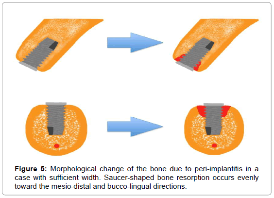
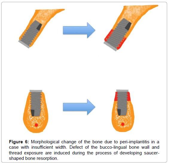
Fransson et al. [12] reported that peri-implantitis was found in about 40% of implants in a 5 to 23 year observation period. As for regions of onset, the rate in the maxillary molar region was as low as 30% and 52% in the mandibular molar region. They reported that implants placed in the anterior region were vulnerable regardless of the failure pattern. The present study resulted in very frequent onsets molar regions in the maxilla and mandibular where plaque control is difficult, similar to that in natural teeth. The different results were found possibly because Fransson et al. studied the peri-implantitis rate in total implants including healthy implants, the rate of implantsupported fixed complete dentures (FCD) without remaining tooth was remarkably high (74%), and bone loss in 3 threads or more and PPD of 6 mm or more were classified as peri-implantitis [16]. The presence of BOP, suppuration, and peri-implant bone resorption found by radiographic evaluation are listed as clinical diagnosis of peri-implantitis. Radiographic evaluation is indispensable for a definite diagnosis, among others, although the use of panoramic tomography and intra-oral radiograph, which are broadly used in clinical settings, does not allow the evaluation of the bucco-palatal bone levels, but only for the observation of mesio-distal bone absorption. Furthermore, the evaluation with CT or cone beam radiography allows no accurate evaluation because of the metal artifacts from implant body and superstructure. Isidor et al. reported the development of the saucershaped bone resorption in the implant in the plaque group [13]. However, it is believed that the saucer-shaped bone resorption occurs in cases with large buccolingually-wide rigde, while the decent lingual/ palatal thread exposure of the implant surface occurs during the development process of saucer-shaped bone resorption in cases with a narrow ridge (Figure 5 and 6). Thus, particularly, the buccopalatal bone resorption even with light or no mesiodistal bone resorption or sometimes severe bone resorptions were frequently observed in the cases with a narrow pyramid-shape ridge. Moreover, DeSmet et al. reported that the conventional radiograph included the inability to monitor both of the facial and lingual/palatal bone levels, the low sensitivity for the detection of early bone changes, and the underestimation of bone loss around implants [11]. In the present study, the implant sites with BOP were examined to observe a number of BOP in the buccal sites of mandibular molar regions and the bucco-palatal sites of maxillary molar regions. Possible reasons for the frequent BOP are narrow rigde that may be found in cases with the mandibular molar region BOP often inducing buccal thread exposure at the time of implantation surgery, great buccolingual thread exposure that tends to occur when saucershaped bone resorption occurs in cases with a narrow bone, and small immobilized mucosa. Problems in buccal immobilized mucosa for the maxillary molars and palatal mucosa thicker than other mucosa with little changes in mucosal thickness in response to placement of upper structure were considered vulnerability to inflammation. These facts also suggest that dental radiography is not always useful in the early diagnosis of peri-implantitis. We would like to continue to develop the measurament that allows the diagnosis of buccal and lingual/palatal bone resorption and its severity other than radiography in the future.
Proposal of Decision Tree for the Diagnosis and Management of Peri-implant Bone Loss
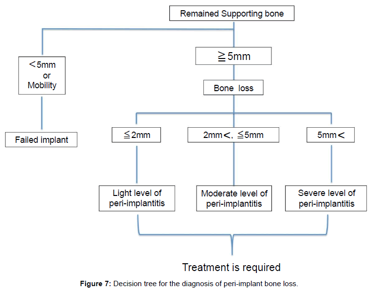
Peri-implantitis is one of the main concerns in implant therapy. The management of peri-implant bone loss is still challenging because of its difficulty and unpredictablity. To manage peri-implant diseases properly, a decision tree that can be used as a guide to assist clinicians in dealing with this increasing challenge was developed. To treat this problem properly, clinicians have to identify the extent of bone loss at the first. The more bone resorption increases, the more difficult the replacement of implants will be due to the anatomical problems. Remaining bone quantity associated with the implants under the continuous bone resorption should also be considered because a number of reports on the prognosis for short implants have been published, such as a review by Renouard reporting that the use of implants with a coarse surface resulted in high success rates with 94.6% to 99.4% survival rates of implants with lengths less than 10 mm [14]. Furthermore, a review by Telleman et al. reported that the survival rate of short implants with 5 to 9.5 mm length was 93.1% to 98.6% in partial failure patients [15]. Thus, the decision tree was developed by defining 5 mm of remaining bone length as the criteria, because we consider it is appropriate that the decision whether clinicians has to remove the implant based on the premise of re-implantation or to conduct conservative therapy should be based on the remaining bone height (Figure 7). In such situations as either remaining bone height with less than 5 mm or the movement of implants, the re-insertion of implants with the bone augmentation following the removal of the implant and the site development is highly recommended, regardless of implant length. Although this condition can also be treated with guided bone regeneration, according to the literature, it is very difficult to regain osseointegration. In regard to implants with remaining bone height with more than 5 mm, it is recommended examining the amount of bone loss. Also, it is suggested to diagnose the bone loss with less than 2 mm as light, with between 2 mm and 5 mm as moderate, and more than 5 mm as severe. Renvert et al. stated in their review, “In peri-implantitis lesions non-surgical therapy was not found to be effective [16]. Adjunctive chlorhexidine application had only limited effects on clinical and microbiological parameters. However, adjunctive local or systemic antibiotics were shown to reduce bleeding on probing and probing depths. Minor beneficial effects of laser therapy on peri-implantitis have been shown; this approach needs to be further evaluated.” Thus, surgical treatment is considered necessary in current therapies even in mild cases. Implant surface decontamination and regenerative procedures are considered important in surgical treatment. However, there are a variety of different procedures with different success rates without a definitely established therapeutic procedure [17]. Thus, our decision tree involves no therapy. Our study will clarify such issues in the future.
References
- van Steenberghe D, Quirynen M, Naert I (1999) Survival and success rates with oral endosseous implants. Proccedings of the 3rd European Workshop on Periodontology. Berlin,Quintessence Books.
- Roos-Jansåker A-M, Lindahl C, Renvert H, Renvert S (2006) Nine- to fourteen-year follow-up of implant treatment. Part II: presence of peri-implant lesions. Journal of Clinical Periodontology 33: 290–295.
- Quirynen M, De Soete M, van Steenberghe D (2002) Infectious risks for oral implants: a review of the literature. Clin Oral Implants Res 13: 1-19.
- Shibli JA, Marcantonio E, d'Avila S, Guastaldi AC, Marcantonio E Jr (2005) Analysis of failed commercially pure titanium dental implants: a scanning electron microscopy and energy-dispersive spectrometer x-ray study. J Periodontol 76: 1092-1099.
- Mombelli A (1999) Prevention and therapy of periimplant infections. Proccedings of the 3rd European Workshop on Periodontology. Berlin, Quintessence Books.
- Lindhe J, Meyle J; Group D of European Workshop on Periodontology (2008) Peri-implant diseases: Consensus Report of the Sixth European Workshop on Periodontology. J Clin Periodontol 35: 282-285.
- Zitzmann NU, Berglundh T (2008) Definition and prevalence of peri-implant diseases. J Clin Periodontol 35: 286-291.
- Heitz-Mayfield LJ (2008) Peri-implant diseases: diagnosis and risk indicators. J Clin Periodontol 35: 292-304.
- Kullman L, Al-Asfour A, Zetterqvist L, Andersson L (2007) Comparison of radiographic bone height assessments in panoramic and intraoral radiographs of implant patients. Int J Oral Maxillofac Implants 22: 96-100.
- Bräegger U, Pasquali L, Weber H, Kornman KS (1989) Computer-assisted densitometric image analysis (CADIA) for the assessment of alveolar bone density changes in furcations. J Clin Periodontol 16: 46-52.
- De Smet E, Jacobs R, Gijbels F, Naert I (2002) The accuracy and reliability of radiographic methods for the assessment of marginal bone level around oral implants. Dentomaxillofac Radiol 31: 176-181.
- Fransson C, Wennström J, Tomasi C, Berglundh T (2009) Extent of peri-implantitis-associated bone loss. J Clin Periodontol 36: 357-363.
- Isidor F (1997) Histological evaluation of peri-implant bone at implants subjected to occlusal overload or plaque accumulation. Clin Oral Implants Res 8: 1-9.
- Renouard F, Nisand D (2006) Impact of implant length and diameter on survival rates. Clin Oral Implants Res 17 Suppl 2: 35-51.
- Telleman G, Raghoebar GM, Vissink A, den Hartog L, Huddleston Slater JJ, et al. (2011) A systematic review of the prognosis of short (<10 mm) dental implants placed in the partially edentulous patient. J Clin Periodontol 38: 667-676.
- Renvert S, Roos-Jansåker AM, Claffey N (2008) Non-surgical treatment of peri-implant mucositis and peri-implantitis: a literature review. J Clin Periodontol 35: 305-315.
- Claffey N, Clarke E, Polyzois I, Renvert S (2008) Surgical treatment of peri-implantitis. J Clin Periodontol 35: 316-332.
Relevant Topics
- Advanced Bleeding Gums
- Advanced Receeding Gums
- Bleeding Gums
- Children’s Oral Health
- Coronal Fracture
- Dental Anestheia and Sedation
- Dental Plaque
- Dental Radiology
- Dentistry and Diabetes
- Fluoride Treatments
- Gum Cancer
- Gum Infection
- Occlusal Splint
- Oral and Maxillofacial Pathology
- Oral Hygiene
- Oral Hygiene Blogs
- Oral Hygiene Case Reports
- Oral Hygiene Practice
- Oral Leukoplakia
- Oral Microbiome
- Oral Rehydration
- Oral Surgery Special Issue
- Orthodontistry
- Periodontal Disease Management
- Periodontistry
- Root Canal Treatment
- Tele-Dentistry
Recommended Journals
Article Tools
Article Usage
- Total views: 16275
- [From(publication date):
September-2014 - Aug 16, 2025] - Breakdown by view type
- HTML page views : 11602
- PDF downloads : 4673

