Make the best use of Scientific Research and information from our 700+ peer reviewed, Open Access Journals that operates with the help of 50,000+ Editorial Board Members and esteemed reviewers and 1000+ Scientific associations in Medical, Clinical, Pharmaceutical, Engineering, Technology and Management Fields.
Meet Inspiring Speakers and Experts at our 3000+ Global Conferenceseries Events with over 600+ Conferences, 1200+ Symposiums and 1200+ Workshops on Medical, Pharma, Engineering, Science, Technology and Business
Editorial Open Access
Parallel Excitation in Ultrahigh Field Human MR Imaging and MultiChannel Transmit System
| Xiaoliang Zhang1,2,3* and Yong Pang1 | |
| 1Department of Radiology and Biomedical Imaging, University of California San Francisco, San Francisco, CA, USA | |
| 2UCSF/UC Berkeley Joint Graduate Group in Bioengineering, San Francisco & Berkeley, CA, USA | |
| 3California Institute for Quantitative Biosciences (QB3), University of California, San Francisco, CA, USA | |
| Corresponding Author : | Xiaoliang Zhang Department of Radiology and Biomedical Imaging University of California San Francisco San Francisco, CA 94158, USA E-mail: xiaoliang.zhang@ucsf.edu |
| Received April 26, 2012; Accepted April 28, 2012; Published April 30, 2012 | |
| Citation: Zhang X, Pang Y (2012) Parallel Excitation in Ultrahigh Field Human MR Imaging and Multi-Channel Transmit System. OMICS J Radiology. 1:e110. doi: 10.4172/2167-79641000e110 | |
| Copyright: © 2012 Zhang X, et al. This is an open-access article distributed under the terms of the Creative Commons Attribution License, which permits unrestricted use, distribution, and reproduction in any medium, provided the original author and source are credited. | |
Visit for more related articles at Journal of Radiology
| Editorial |
| Parallel excitation [1,2] with a multi-element Radio Frequency (RF) transceiver array [3-9] as a contemporary methodology has been advocated for human MR imaging at ultrahigh magnetic fields (7 Tesla and above). In ultrahigh field MRI, the required high operating frequency and thus shortened wavelength of radio frequency waves creates a complex wave behavior and increased phase variation of RF magnetic fields (i.e. B1 fields) in conductive and high dielectric biolog- ical samples, such as human body, resulting in inhomogeneous image distribution. The inhomogeneous image distribution consequently leads to difficulties in quantifying the MR signal intensity. With inde- pendent phase and amplitude control of each channel of a transceiver array, parallel excitation can be applied to perform B1 shimming to obtain uniform B1 distribution. In MR safety aspect, RF power re- quired to excite the spins increases dramatically at ultrahigh fields compared with that at lower fields, e.g. 1.5T. The high RF excitation power results in high Specific Absorption Rate (SAR) in human body, ultimately increases tissue heating during MRI. It is demonstrated that by using the parallel excitation method, the RF excitation pro- file can be optimized, providing in a significantly reduced SAR and therefore safer MRI at ultrahigh fields. In fact, the emerging method of parallel excitation has become essential for ultrahigh field MRI in addressing B1 in homogeneity, increased SAR and tissue heating. |
| Additionally, parallel excitation with a multi-element RF trans- ceiver array has opened a new avenue to selective excitation in MR imaging, providing a fast and efficient approach to perform selective excitation [1,2]. Conventionally a single RF pulse is used in MRI to perform slice selective or multidimensional spatial selective exci- tation by exciting the nuclei in the area of interest and limiting the electromagnetic signal emitted from imaging object within spatially restricted areas [10-16]. This often requires homogeneous RF field to ensure excitation accuracy. However, as described above it is techni- cally challenging to achieve homogeneous B1 fields with the increase of the magnetic field strength, where the dielectric resonance [17] and the conductivity effect of high dielectric and conductive biological samples [18,19] lead to enlarged B1 field variation [20] even with an intrinsically homogeneous volume coil [4,21,22,23]. This effect be- comes more pronounced at ultrahigh field such as 7 Tesla (7T) due to the shortened wavelength of radio frequency (RF) wave [24]. In con- ventional selective excitations, the pulse width of the required multi- dimensional RF pulses is usually long, resulting in a long excitation time, especially in applications where a large excitation Field Of View (FOV) is involved. Such long excitation could potentially exacerbate the high SAR at ultrahigh fields. Although special k-space trajectory such as spiral trajectory and iterative pulse design method have been developed to reduce the length of multidimensional RF pulse [25-27], multiple pulse parallel excitation method adopted from parallel im- aging is able to significantly reduce the excitation time, providing a whole new approach to selective excitation and more capabilities than conventional single RF pulse excitation [1-3,5,6,8,9,28-30]. Parallel excitation RF pulses were originally designed to shorten the duration of multidimensional spatially selective excitation [1,2,28,31-35]. This capability has been used for both small tip-angle excitation and large tip-angle multidimensional RF pulses [36-38]. By using the mul- tichannel parallel excitation, the pulse width for multidimensional spatial selective excitation can be dramatically reduced. Parallel ex- citation can also take advantage of independent control of each RF pulse to reduce the RF power and minimize the SAR [2,39-48] dur- ing the multidimensional spatial selective excitation. Furthermore, by using multiple independent RF pulses, the phase and amplitude of each pulse can be adjusted to manipulate the excitation profiles to achieve the desired RF field in conductive and high dielectric biologi- cal sample [44,49-52]. These capabilities, achieved by utilizing the ex- tra degree of freedom from multiple RF pulse excitation, can provide much more advantages over conventional single RF transmission. Ap- parently, to design a practically optimal RF pulses for selective excita- tion using parallel excitation, it is necessary to not only optimize the excitation profile homogeneity, but also minimize the peak RF power to reduce the SAR [27,53,54] in order to ensure the safety during MR examinations. |
| To enable parallel excitation for B1 shimming, SAR optimization and multiple RF pulse excitation, an MR scanner must be equipped with multi-channel transmitters which can independently control the amplitude and phase of RF pulse on each channel. However, in cur- rent system setup, most existing MR scanners used in research insti- tutions and clinical settings are not equipped with the multi-channel transmitters and thus are not capable of implementing this emerging concept of parallel excitation for B1 shimming, SAR optimization and fast selective excitation. In recent years, some parallel transmit sys- tems have been developed to allow parallel excitation applications on existing MR scanners [55-57]. These designs can provide 100 Watts power (or higher, depending on the type of amplifiers used) for each RF excitation channel and can be used for simultaneously multiple RF pulse excitations on commercial MR scanners. |
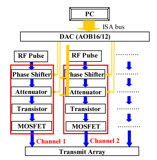
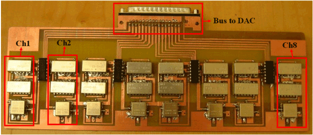
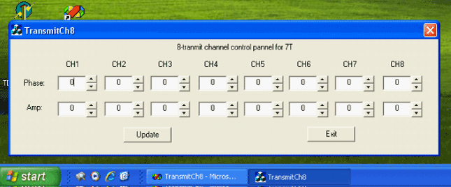
| In practice, it is desired to have a multi-channel transmitter sys- tem which is easy to be integrated to the existing MR scanners. In this research endeavor, a PC controlled 8-channel transmit circuit with independent phase and amplitude for each channel for 7T MR scan- ner has been proposed [57]. In this design, the phase and amplitude of each channel are adjusted by voltage control phase shifter and attenuator respectively. Both control voltages are communicated through a PC via a 16-channel Digital Analog Converter (DAC). The input of this circuit can be the RF pulse generated by the signal generator of the MR spectrometer. Through this phase and amplitude control circuit, the output signal of each channel can be amplified for spin excitation by RF amplifiers which could be either regular RF amplifiers or on-coil MOSFET amplifiers [58,59]. Figure 1 shows the block diagram of this multi-channel transmitter circuit design. The PC is utilized to send out digital voltages which determine the phase shift and amplitude change of each channel. The circuit board of the 8-channel transmit system is shown in Figure 2. A Graphic User Interface (GUI) for PC was also developed to facilitate the control of the output voltages of the DAC (Figure 3). |
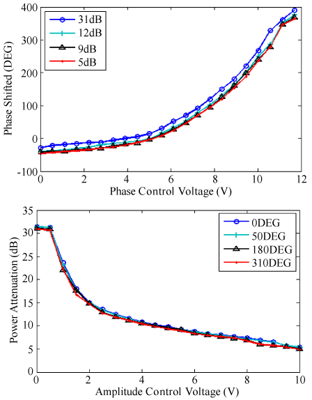
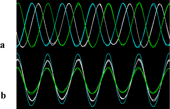
| We utilized the benchmark signal to test its performance. A 298MHz sine waveform was output to the 8-channel transmit circuit and an oscilloscope was used to display the output waveforms from the circuit. The phases and amplitude varying with the control voltages were plotted in Figure 4, demonstrating the sufficient dynamic range of the design for MR applications. Several samples of the pulse waveforms with different phases and amplitudes are shown in Figure 5. By synchronizing the clock signal to the excitation signal of MRI scanner, this design should be readily integrated to existing MR scanner. |
| This PC controlled 8-channel transmit circuit design provides a comparatively simple way to enable parallel excitation applications for the existing MR scanners that are not equipped with multi-channel transmitter systems. With this, parallel excitation for B1 shimming, SAR optimization and fast selective excitation can be performed. |
| Acknowledgements |
| This work was partially supported by NIH grants EB004453 and EB008699, and a QB3 Research Award. |
References |
|
Figures at a glance
 |
 |
 |
 |
 |
| Figure 1 | Figure 2 | Figure 3 | Figure 4 | Figure 5 |
Post your comment
Relevant Topics
- Abdominal Radiology
- AI in Radiology
- Breast Imaging
- Cardiovascular Radiology
- Chest Radiology
- Clinical Radiology
- CT Imaging
- Diagnostic Radiology
- Emergency Radiology
- Fluoroscopy Radiology
- General Radiology
- Genitourinary Radiology
- Interventional Radiology Techniques
- Mammography
- Minimal Invasive surgery
- Musculoskeletal Radiology
- Neuroradiology
- Neuroradiology Advances
- Oral and Maxillofacial Radiology
- Radiography
- Radiology Imaging
- Surgical Radiology
- Tele Radiology
- Therapeutic Radiology
Recommended Journals
Article Tools
Article Usage
- Total views: 14422
- [From(publication date):
May-2012 - Aug 16, 2025] - Breakdown by view type
- HTML page views : 9780
- PDF downloads : 4642
