Review Article Open Access
256-Slice Coronary Computed Tomography Angiography Using Low Tube Voltage of 100 KV
| Bhojraj Sharma* | |
| Gandaki Medical College Teaching Hospital and Research Centre, Nepal | |
| Corresponding Author : | Bhojraj Sharma Lecturer, Gandaki Medical College Teaching Hospital and Research Centre Medical Imaging and Nuclear Medicine (Radiology), Lakenath, Kaski, Nepal Tel: +9779856044660 E-mail: bhojrajsharma2@gmail.com |
| Received January 18, 2014; Accepted February 16, 2015; Published February 20, 2015 | |
| Citation: Sharma B (2015) 256-Slice Coronary Computed Tomography Angiography Using Low Tube Voltage of 100 KV. OMICS J Radiol 4:180. doi: 10.4172/2167-7964.1000180 | |
| Copyright: © 2015 Sharma B. This is an open-access article distributed under the terms of the Creative Commons Attribution License, which permits unrestricted use, distribution, and reproduction in any medium, provided the original author and source are credited. | |
Visit for more related articles at Journal of Radiology
Abstract
Purpose: To evaluate the image quality and radiation dose of 100 kV with 1000 mAs retrospective electrocardiography (ECG)-gated CCTA protocol, compared to the standard protocol of 120 kV with 800 mAs retrospective ECG-gated CCTA. Material and Methods: We divided 70 patients into two, a reduced dose group with 35 patients (18 M, 17 F; Mean age 56.94 ± 11.51 years) were examined by 100-kV with 1000 mAs retrospective ECG-gated CCTA, and another as a standard group of 35 patients (21, 14 F; Mean age 54.03 ± 9.81 years) were examined by 120-kV with 800 mAs retrospective ECG-gated CCTA. The two blinded radiologists analyzed the image quality of the coronary arteries independently and they accessed subjective and objective image quality. The radiation dose was also measured as effective radiation dose [ED] and was calculated using CT dose volume index [CTDIvol.], dose-length product [DLP] and conversion coefficient for chest (conversion factor k=0.014 mSv mGy-1cm-1). Results: Although the objective image quality of the 100-kV with 1000 mAs was significantly better than the 120- kV with 800 mAs (mean SNR, 36.65 ± 2.95 vs. 33.47 ± 3.86, P<0.0001; mean CNR, 34.27 ± 2.92 vs. 30.62 ± 3.90, P<0.0001). There was no significant variation in the subjective image quality between two groups (mean image score, 4.54 ± 0.37 vs. 4.56 ± 0.25 for radiologist 1, P = 0.781; 4.52 ± 0.25 vs. 4.56 ± 0.25 for radiologist 2, P=0.486). The radiation dose was found to be reduced by 28% with the 100-kV/1000 mAs protocol than with the 120-kV/800 mAs retrospective ECG-gated CCTA (7.87 ± 0.59 vs. 10.95 ± 1.67 mSv, P<0.0001). Conclusion: The protocol of low tube voltage CCTA using 100 kV/1000 mAs retrospective ECG-gated shows significant reduction of the radiation dose without disturbing the subjective image quality of CCTA.
| Keywords |
| Coronary CT angiography; Radiation dose; Image quality |
| Introduction |
| Coronary CTA has good diagnostic performance for detecting CAD as it can provide important information in diagnosis of atherosclerosis [1]. The latest advancements such as increase in speed of gantry rotation, multidetector array and dual-source detectors, increase its capability to visualize anatomy of coronary arteries noninvasively and to identify location, magnitude and composition of coronary atherosclerosis. It has been shown that CCTA has high diagnostic accuracy for obstructive CAD in comparison to invasive coronary angiography. It helps in classification between calcified and non- calcified CAD which shows its value of prognosis and in early detection for risk stratification. CCTA provides stable image quality and ability to diagnose coronary artery stenosis with a high negative predictive value that seems very important in clinical applications [2]. There is high sensitivity (96- 99%) and specificity (88-91%) for diagnosing coronary artery disease by Coronary CT angiography. Although it has a good diagnostic performance in detecting coronary artery disease, the radiation dose may be high that has risk of cancers. |
| Materials and Methods |
| Study population |
| Seventy consecutive patients were enrolled in this prospective study from September 2012 to February 2013. All the included patients signed informed consent to be in study and the study was approved by institutional review board. The inclusion criteria used in this study was a normal body mass index, ranging between BMI of 18.5 and 25 kg/m. The exclusion criteria in this study exclude patients with serum creatinine level more than 1.5 mg/dl and previous adverse reaction to iodine contrast. All patients had suspected or known CAD investigated for clinical indications. The indications were according to current guidelines and recommendations [3]. The patients were randomly allocated into two different CTCA protocols. Thirty-five patients were examined with reduced dose protocol of 100 kV with 1000 mAs retrospective ECG-gated CCTA. The other thirty-five patients were examined by using standard dose protocol of 120-kV with 800 mAs retrospective ECG-gated CTCA and described as the standard group. |
| Preparation of the patient |
| The standard protocol in our department for coronary CT angiography was followed for the examination. The patients were explained about the examination protocol which includes information to avoid caffeinated drinks on the day of scanning, to remain fasting for at least 2 hours ahead of the examination, and measurement of the heart rate and blood pressure 1 hour before starting the scan and informed consent was taken from all the patients. Then the vascular access by using 20-gauge retractive safety IV catheter was obtained in the veins of the dorsal right hand (i.e. portion of the cephalic vein near the radial styloid process). All the patients were instructed about breath holding technique before starting the examination. |
| Image acquisition |
| All the scanning examinations were done on a 256-slice Multidetector CT scanner (Brilliance iCT, Philips Healthcare, Cleaveland, Ohio, USA). It had a detector collimation of 128 × 0.625 mm with double z-sampling. This scanner had a spatial resolution of 0.625 mm, 0.27 sec gantry rotation time and temporal resolution of 135 msec. The temporal resolution of this machine had been further improved by using advanced cardiac adaptive multi-cycle reconstruction algorithms which combined data from consecutive cardiac cycles [4]. The scanner of Philips iCT Brilliance offers 80 mm of z-axis coverage during a single gantry rotation. The 120 kV with an effective tube current-rotation time product of 800 mAs was applied for the standard group. We applied different approach to reduce the radiation dose in another group. The tube voltage was reduced to 100 kV while the tube current-rotation time product was increased to 1000 mAs, in comparison to the standard CTCA protocol (Table 1). |
| Contrast material |
| The protocol of application of contrast agent was similar in both groups. The contrast material named Xenetix 350 (Lobitridol, Guerbet Asia Pacific, Shanghai, China) at 1 ml/kg body weight was applied for the examination. Xenetix was first produced in 1994. It is a non-ionic, low osmolality iodinated contrast agent which can be used for wholebody imaging, conventional cardiovascular imaging and intravascular imaging. Its efficacy was confirmed during evaluation of more than 163,700 patients in 3 German surveys [5-7]. |
| Injection protocol |
| Depending upon BMI of the patients, volume of the contrast agent was set. It is approximately 70-80 ml of Xenetix 350 given at 5.0 ml/s rate followed by 50 ml of saline chaser (9% normal saline) at 5.0 ml/s by using automated dual-syringe injector (Empower CTA dualsyringe injector) with the pressure limit set at 300 PSI. The application of contrast agent was controlled by bolus tracking method kept in the descending aorta with signal attenuation threshold of 120 HU. |
| Acquisition protocol |
| CT scanning was performed in a cranio-caudal direction from the carina level to the bottom of the heart (level of diaphragm). The CT data were reconstructed with a slice thickness of 0.90 mm with a reconstruction increment of 0.45 mm using Adaptive Filter. In this study, all scans were performed using retrospective ECG-gating method with a 120 kV tube voltage, 0.27 s of rotation time, effective tube current-rotation time product of 800 mAs, a pitch factor of 0.16 and a fixed detector collimation of 128 × 0.625 mm in standard group and 100 kV with 1000 mAs in another reduced dose group. Advanced cardiac adaptive multi-cycle reconstruction algorithms that combine data from cardiac cycle was also used to further improve the standard temporal resolution of 135 ms. The field-of-view (FOV) obtained was 250 mm. The delivery of contrast agent was controlled by automatic bolus tracking (Bolus Pro, Philips Healthcare, Cleveland, OH, USA) with defining a region of interest (ROI) in the center of descending aorta at aortic root level. The initiation of the scan was after a post-threshold delay of 6 sec after the signal attenuation reached a predetermined threshold of 120 HU in the descending aorta (Table 1). |
| Image reconstruction |
| For the reconstruction of image after scanning and evaluation of the quality of images scanned, a dedicated workstation (Extended Brilliance Workspace [EBW] Version V4.5.2.40007, Philips Healthcare, Cleveland, Ohio, USA) was used. Xres Standard filter (XCB, Philips Healthcare, Cleveland, Ohio, USA) was used for the purpose of image reconstruction with axial images, coronal, sagittal and curved multiplanar reformations. Raw data reconstruction was obtained by using 0.9 mm slices at 0.45 mm intervals with different intervals of the R-R phase. Straightened MIP reformations were obtained by slice thickness of 3.0 mm. The more optimal phase for reconstruction was selected to reduce the effect in case of motion artifact in a vessel on review at the beginning. |
| Post processing and image analysis |
| Two radiologists, blinded to the division of patients groups, analyzed image quality after optimal selection of R-R reconstruction phases. Decisions were made on consensus reading. Image quality analysis was done on axial slices, curved MPR and straightened MIP projections. The two radiologists provided both subjective and objective analysis of the image quality independently. For subjective analysis of the image quality, the criteria used by both radiologists was overall appearance of the vessel, internal and external wall definition, degree of motion artifact, differentiation between calcified and noncalcified plaque and vessel lumen. They analyzed the image quality at the same time through a consensus approach using a 5-point scoring scale: 1 (excellent without artifacts in all arteries and clear delineation of vessels); 2(good with minor artifacts or mild blurring in at least one main vessel); 3 (fair with moderate artifacts or moderate blurring without discontinuity of vessel); 4 (poor with severe artifacts, doubling or discontinuity of at least one vessel); or 5 (non-diagnostic). For the evaluation of objective image quality, the CT image attenuation value was measured in Hounsfield Units (HU) and image noise was measured as standard deviation (SD) in the region of interest (ROI) with area of 1 cm in ascending aorta at level of LM origin on axial images [8]. |
| Effective dose calculation |
| The volume computed tomography dose index (CTDIvol) was obtained for each patient from the EBW. The dose-length product (DLP) was calculated as product of CTDIvol and the craniocaudal distance of scanned part (scan length in cm) in each patient separately. The estimated effective dose in mSv per patient was calculated by product of the DLP and a conversion coefficient for chest (conversion factor k=0.014 mSv mGy cm) [9]. |
| Statistical analysis |
| We used 120-kV protocol as the standard CTCA protocol. All of the parameters of reduced tube voltage protocol of 100 kV were compared with that of the standard protocol. In the beginning, we entered all the variables and data in Microsoft Excel software. All statistical analyses were done by using the Statistical Package for the Social Sciences (SPSS) for Windows 32 bit edition, version 21.0.0.0 (IBM Corporation, 2012). We transferred all variables and data into SPSS software from Excel. The quantitative or continuous variables were defined as mean ± standard deviation. The categorical variables were defined as frequencies or percentages. We considered associations significant at P values<0.05. We applied Student independent t-test to compare between the means of continuous variables. While doing comparison between the categorical variables, we used chi-square test. To assess inter-observer reproducibility of the image quality by the subjective method between two observers, we used the interclass correlation test. A Cronbach’s α<0.70 will indicate a strong correlation between them, value between 0.40 and 0.70 will indicate moderate correlation and value less than 0.40 will indicate weak correlation between them. |
| Results |
| Demographic study of patients |
| The two divided protocols showed reduced dose group included (18 Males, 17 Females; Mean age 56.94 ± 11.51 years) patients and standard group included (21 Males, 14 Females; Mean age 54.03 ± 9.81 years) patients. There was no significant variability between two groups of 100 kV and 120 kV protocols in age (56.94 ± 11.51 vs. 54.03 ± 9.81, P=0.258), sex (male/female distribution; 18/17 vs. 21/14, P = 0.470). The BMI (22.39 ± 1.60 vs. 22.68 ± 1.54, P=0.455), and heart rate (81.14 ± 12.23 vs. 80.63 ± 15.8, P = 0.879) also showed no significant variability between two protocols (Table 2). |
| Image quality of coronary arteries |
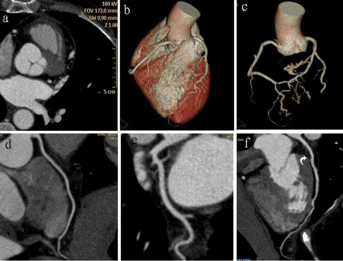
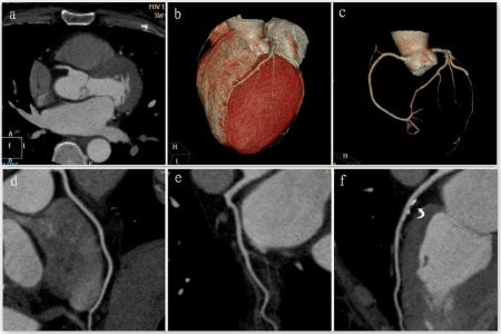
| The mean image noise, the attenuation of the proximal RCA, LM, LAD, LCX, ascending aorta and the attenuation of chest wall muscle were different according to different tube voltage protocols of CCTA. The mean image noise of 100 kV protocol was 1.19 times higher than 120 kV protocol (12.80 ± 0.45 vs. 10.74 ± 0.45 respectively; P-value<0.0001). The attenuation value of RCA is 1.30 times higher in reduced dose protocol than in standard protocol (468.61 ± 33.82 vs. 360.03 ± 38.79 respectively; P-value<0.0001). We found the attenuation value in LM with 100 kV is 1.31 times higher than in 120 kV protocol (467.58 ± 34.32 vs. 358.20 ± 38.14 respectively; P-value <0.0001). In reduced dose group, the attenuation value in LAD is 1.31 times higher than in 120 kV protocol (465.58 ± 34.57 vs. 354.59 ± 38.32 respectively; P-value<0.0001). The attenuation value in LCX with 100 kV is 1.32 times higher than in 120 kV protocol (465.38 ± 33.43 vs. 353.74 ± 38.00 respectively; P-value<0.0001). The attenuation value in ascending aorta with 100 kV protocol is 1.3 times higher than in 120 kV protocol (476.42 ± 33.19 vs. 367.39 ± 40.41 respectively; P-value<0.0001). The mean attenuation value with 100 kV is 1.31 times higher than in 120 kV protocol (468.72 ± 33.75 vs. 358.79 ± 38.63 respectively; P- value <0.0001) (Table 3, Figures 1 and 2). |
| Objective image quality |
| The mean SNR was 1.1 times higher in 100 kV than in 120 kV (36.65 ± 2.95 vs. 33.47 ± 3.86 respectively; P<0.0001). The mean CNR was 1.12 times higher than 120 kV (34.27 ± 2.92 vs. 30.62 ± 3.90 respectively; P<0.0001) as shown in Table 3. |
| Subjective image quality |
| The subjective image quality between two protocols (100 kV protocol and standard protocol of 120 kV) by 2 radiologists, the score of each main vessels, sum of four vessels and mean image score of these vessels was not significantly different between these two protocols. The different scores in 100 kV and 120 kV by radiologist 1 was for RCA (4.6 ± 0.6 vs. 4.6 ± 0.6 respectively; P-value = 0.809), for LM (4.9 ± 0.3 vs. 4.7 ± 0.5 respectively; P = 0.070), for LAD (4.4 ± 0.6 vs. 4.6 ± 0.5 respectively; P=0.435), for LCX (4.2 ± 0.5 vs. 4.4 ± 0.6 respectively; P=0.113). For Radiologist 2, the different scores in 100 kV and 120 kV was for RCA (4.6 ± 0.6 vs. 4.6 ± 0.6 respectively; P-value = 1.000), for LM (4.8 ± 0.4 vs. 4.6 ± 0.5 respectively; P = 0.166), for LAD (4.4 ± 0.6 vs. 4.6 ± 0.5 respectively; P=0.251), for LCX (4.3 ± 0.6 vs. 4.4 ± 0.6 respectively; P=0.379). The mean image scores in reduced and standard protocols by radiologist 1 and radiologist 2 was 4.5 ± 0.4 vs. 4.6 ± 0.2; P = 0.663 and 4.5 ± 0.3 vs. 4.5 ± 0.3; P = 0.860 respectively. The interobserver reproducibility was moderate for all four vessels (Cronbach’s α 0.568 for RCA, 0.609 for LMA, 0.560 for LAD and 0.579 for LCX). The subjective image quality score obtained from two radiologists in two protocols were summarized in Table 4. |
| Estimated radiation dose |
| The radiation dose estimated in two different protocols was summarized in Table 4. There was no any significant difference between the scan range of 100 kV protocol and the standard protocol of 120 kV (14.11 ± 1.05 vs. 14.71 ± 2.25 respectively; P=0.155). The reduction of tube voltage in 100 kV protocol reduced all three parameters of CTDIvol (39.9 ± 0.00 vs. 53.2 ± 0.00; P<0.0001), DLP (562.94 ± 42.12 vs. 782.64 ± 119.49; P<0.0001), and ED (7.87 ± 0.59 vs. 10.95 ± 1.67; P<0.0001) in comparison to standard protocol of using 120 kV during CCTA respectively. In comparison between them, there was marked reduction of ED by 28% in 100 kV protocols (Table 5). |
| Discussion |
| In this prospective study, there was no significant variability between two groups of 100 kV and 120 kV protocols in age, sex, BMI and heart rate. There was an estimated radiation dose reduction by 28% while using 100 kV protocol CCTA than in 120 kV standard protocol CCTA. Although there was better objective image quality in 100 kV protocol CCTA, there was no any significant difference between the subjective image quality obtained by 2 radiologists in two different protocols of 100 kV and 120 kV. The mean image noise, the attenuation of the proximal RCA, LM, LAD, LCX and the ascending aorta was higher in 100 kV reduced dose protocol in comparison to 120 kV standard dose protocol. |
| The use of CCTA is very helpful to diagnose coronary artery disease. The meta- analyses study of CCTA showed high sensitivity (96%-99%) and specificity (88%-91%) for diagnosing Coronary artery disease [10]. But there is increased risk of cancer due to high radiation dose. There may be lifetime cancer incidence up to 0.2% and 0.7% for patients doing 16 slice CCTA and 64 slice CT scanners respectively and 0.4% for lung and breast cancer has been reported [11]. This current study found that the radiation estimated dose can be reduced by lowering tube voltage during CCTA. It can be done reducing tube voltage from 120 kV to 100 kV in patients of BMI below 25 kg/m2 without degradation of the subjective and objective image quality obtained by the scanning. Even though the two different observes studied the subjective image quality of the two groups, there is no much dissimilarity between the score obtained by them. The interclass correlation test indicates the inter-observer reproducibility was found to be moderate. Although 100 kV reduced dose protocol increased the image noise than 120 kV (12.80 ± 0.45 vs. 10.74 ± 0.45 respectively; P<0.0001), the objective image quality in 100 kV protocol is better than 120 kV standard doses protocol. Because the mean attenuation of coronary arteries in 100 kV is significantly higher than 120 kV (468.72 ± 33.75 vs. 358.79 ± 38.63 respectively; P<0.0001). This indicates that even we reduce the tube voltage during coronary CTA, the image quality remains same. All three parameters of CTDIvol, DLP and ED were decreased in reduced dose protocol than in standard protocol. The strategy of study was to reduce the estimated radiation dose during CCTA scanning by using low tube voltage protocol with retrospective ECG gated technique. This study concluded that by using low tube voltage protocol of 100 kV instead of using standard 120 kV protocol, we can reduce the radiation dose by 28 percentages in patients with BMI below 25 kg/m2. The subjective image quality was maintained in comparison to the standard protocol by lowering the tube voltage. |
| Weigold et al showed that ED of 11.4 had been obtained during retrospective ECG gated CCTA in 256 slice CT scanner [12]. By using some method, we can reduce the hazards from radiation dose during CCTA. Sebastian Leschka et al found that the radiation dose can be markedly reduced from 8.9 mSv to 6.7 mSv while reducing tube voltage from 120 kV to 100 kV during scanning by dual source CCTA with retrospective ECG gating technique [13]. Paul Stolzmann et al described that reduced tube voltage will reduce 20% dose of contrast material without disturbing the subjective and objective image quality while performing by dual source CCTA [14]. There was reduction of radiation dose by 31% in 64 slice CCTA in non-obese patients in the study performed by Jorg Hausleiter et al. [15]. Bo Ram Jun et al. performed 64 slice CCTA and found that there was an significant reduction in CTDIvol, DLP and ED by 70% while performing with low tube voltage with 80 kV protocol. There was an additional radiation dose reduction with ECG based modulation of tube current by 40% but the noise of images had been increased [16]. Tobias Pflederer et al performed dual source CCTA and got the result that there was significant reduction in radiation dose from 12.7 ± 1.7 mSv to 7.8 ± 2.0 mSv by reducing the tube voltage from 120 kV to 100 kV with ECG pulsing protocol [17]. If there is presence of heavy calcification in coronary arteries, the low tube voltage technique may disturb the diagnostic accuracy of CCTA. With elevated and irregular heart rates, the image quality may hamper. There may be blooming artifact due to dense calcification in the coronary arteries which can lead to overestimation of degree of stenosis. Evaluation of metallic stents can also be limited due to the blooming artifacts from the stent struts. However Michael Lell et al. performed CTCA in 25 patient weighing below and above 100kgs with 100 kv/300 mAs and 120 kV/400 mAs respectively and concluded that ECG-triggered helical CT with high pitch acquisition can yield high and stable image quality with low radiation dose [18]. Gudrun M. Feuchtner et al analyzed patients with retrospective ECG-pulsing spiral 64-slice CCTA and found that the attenuation value of coronary artery was higher in 100 kV group. They showed that 100 kV protocol significantly decreased radiation dose in CCTA with low BMI patients (<25 kg/m2) whereas it showed high image quality [19]. |
| Conclusion |
| In conclusion, with the application of low tube voltage protocol of 100 kV as done in our study, we can reduce the estimated radiation dose by 28% in comparison to the standard protocol of 120 kV protocol during examination of CCTA. There was no any significant difference in the image quality obtained by the subjective method between these two protocols by two radiologists although there was increased in image noise in reduced dose protocol. As there is no degradation of the image quality, we can apply 100 kV protocol as in our study to perform examination of coronary arteries in patients having BMI below 25 kg/ m2 and the BMI should be obtained before the initiation of coronary artery examination. |
References |
|
--
Tables and Figures at a glance
| Table 1 | Table 2 | Table 3 | Table 4 | Table 5 |
Figures at a glance
 |
 |
| Figure 1 | Figure 2 |
Relevant Topics
- Abdominal Radiology
- AI in Radiology
- Breast Imaging
- Cardiovascular Radiology
- Chest Radiology
- Clinical Radiology
- CT Imaging
- Diagnostic Radiology
- Emergency Radiology
- Fluoroscopy Radiology
- General Radiology
- Genitourinary Radiology
- Interventional Radiology Techniques
- Mammography
- Minimal Invasive surgery
- Musculoskeletal Radiology
- Neuroradiology
- Neuroradiology Advances
- Oral and Maxillofacial Radiology
- Radiography
- Radiology Imaging
- Surgical Radiology
- Tele Radiology
- Therapeutic Radiology
Recommended Journals
Article Tools
Article Usage
- Total views: 14420
- [From(publication date):
February-2015 - Aug 28, 2025] - Breakdown by view type
- HTML page views : 9844
- PDF downloads : 4576
