 Spanish
  Chinese
  Russian
  German
  French
  Japanese
  Portuguese
  Hindi
Hindi Our Group organises 3000+ Global Conferenceseries Events every year across USA, Europe & Asia with support from 1000 more scientific Societies and Publishes 700+ Open Access Journals which contains over 50000 eminent personalities, reputed scientists as editorial board members.
Open Access Journals gaining more Readers and Citations
700 Journals and 15,000,000 Readers Each Journal is getting 25,000+ Readers
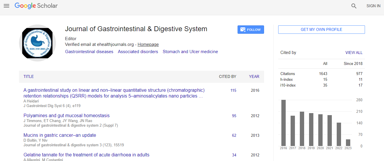
Google Scholar citation report
Citations : 2091
Journal of Gastrointestinal & Digestive System received 2091 citations as per Google Scholar report
Journal of Gastrointestinal & Digestive System peer review process verified at publons

Indexed In
- Index Copernicus
- Google Scholar
- Sherpa Romeo
- Open J Gate
- Genamics JournalSeek
- China National Knowledge Infrastructure (CNKI)
- Electronic Journals Library
- RefSeek
- Hamdard University
- EBSCO A-Z
- OCLC- WorldCat
- SWB online catalog
- Virtual Library of Biology (vifabio)
- Publons
- Geneva Foundation for Medical Education and Research
- Euro Pub
- ICMJE
Useful Links
Recommended Journals
Related Subjects
Share This Page
Incidentally detected space occupying lesions in liver imaging in chronic hepatitis C and the risk of hepatocellular carcinoma
13th Euro-Global Gastroenterology Conference
Saad Al Kaabi, Maneesh Khanna, Madiha E Soofi and Ragesh B Thandassery
Hamad General Hospital, Qatar
Posters & Accepted Abstracts: J Gastrointest Dig Syst
Abstract
Aim: The aim of this study is to evaluate the occurrence and nature of space occupying lesions (SOL) on liver imaging in patients with chronic hepatitis C (CHC) receiving antiviral therapy (AVT). Methods: 1540 patients (from January 2002 to July 2014) with CHC who underwent ultrasound (USG) scan, liver biopsy (LB) and received conventional (pegylated interferon and ribavirin) dual AVT were retrospectively analyzed for the occurrence and nature of SOL prior to initiation of AVT and thereafter. Results: Mean age of patients were 41.9±9.7 years (85% males), predominantly genotype 4 (65%) and genotype 1 (11%). Pretreatment LB showed (Scheuer classification) stage-0 fibrosis (F0) in 1.9%, stage-1 (F1) in 32.9%, stage-2 (F2) in 39.5%, stage-3 (F3) in 19% and stage-4 (F4) in 6.6% patients. Median follow-up was 3.5 years (5390 patient years). Computed Tomography (CT) and Magnetic resonance imaging (MR) scans were performed in 1185 and 560 patients respectively prior to AVT and during follow up. Of the patients with F4 on LB, USG identified cirrhosis in 68%. Of all the patients reported with cirrhosis on USG, F4 was seen in 16.6% and advanced fibrosis (F3 and F4) in 53.4%. Routine pretreatment USG showed fatty liver in 334 (20.8%). Incidental SOL included, cysts in 21 (1.3%), hemangioma in 41 (2.6%) and hypoechoic lesions in 14 (0.9%). CT and/or MR scan did not change diagnosis in most of these SOL, except in two hypoechoic lesions detected pretreatment (one was confirmed as hepatocellular carcinoma (HCC) by CT and another by MR) and one hypoechoic lesion during post AVT follow up (confirmed as HCC in MR). CT and MR identified three and one HCC respectively that were missed on USG against cirrhotic background. Four patients had HCC during pretreatment evaluation and 11 developed new onset HCC during post AVT follow up. The mean alpha fetoprotein (AFP) at diagnosis of new onset HCC was 63.3±1173.8 (vs 16.2±118.1 for non HCC patients, p=0.001). Elevated AFP levels were seen in 81.8% of these patients. Independent pretreatment predictors of new onset HCC were pre-AVT albumin and GGT. Conclusions: Most of the incidental SOL detected in routine pre-AVT USG retained diagnostic consistency with further radiological imaging with CT and MR. A predicting model including pre-AVT albumin and GGT showed high predictive accuracy for development of HCC during post treatment follow up; which could be used as a guide to consider further radiologic evaluation in these incidental SOL.Biography
E-mail: SAADALKAABI@hamad.qa