 Spanish
  Chinese
  Russian
  German
  French
  Japanese
  Portuguese
  Hindi
Hindi Our Group organises 3000+ Global Conferenceseries Events every year across USA, Europe & Asia with support from 1000 more scientific Societies and Publishes 700+ Open Access Journals which contains over 50000 eminent personalities, reputed scientists as editorial board members.
Open Access Journals gaining more Readers and Citations
700 Journals and 15,000,000 Readers Each Journal is getting 25,000+ Readers
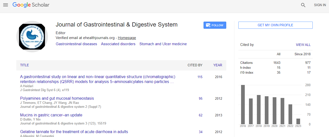
Google Scholar citation report
Citations : 2091
Journal of Gastrointestinal & Digestive System received 2091 citations as per Google Scholar report
Journal of Gastrointestinal & Digestive System peer review process verified at publons

Indexed In
- Index Copernicus
- Google Scholar
- Sherpa Romeo
- Open J Gate
- Genamics JournalSeek
- China National Knowledge Infrastructure (CNKI)
- Electronic Journals Library
- RefSeek
- Hamdard University
- EBSCO A-Z
- OCLC- WorldCat
- SWB online catalog
- Virtual Library of Biology (vifabio)
- Publons
- Geneva Foundation for Medical Education and Research
- Euro Pub
- ICMJE
Useful Links
Recommended Journals
Related Subjects
Share This Page
Parietization of colon following Tuberculous Ascites
14th Euro-Global Gastroenterology Conference
Shailesh Kumar
Dr. Ram Manohar Lohia Hospital, IndiaPost Graduate Institute of Medical Education & Research, India
ScientificTracks Abstracts: J Gastrointest Dig Syst
Abstract
A 46 years old menopausal female presented to surgical OPD with the complaints of recurrent pain abdomen with vomiting and fever off and on. Pt was a treated case of Koch’s abdomen. There was no history of jaundice and other co- morbidities. On examinations, she had tenderness in Right hypochondrium (RHC) on deep palpation. Rest of the parameters were normal. On Investigation, ultrasonography of abdomen revealed multiple gallstones with Normal CBD. Rest of the abdomen and pelvis were normal. Her blood and urinary examinations were within normal limits. X-ray chest revealed features suggestive of healed tuberculosis. Pt was posted for laparoscopic cholecystectomy. After pneumo-peritoneum, 10 mm optical port was placed in periumbilical area. On diagnostic laparoscopy, whole of the colon was densely adhered to the pariety. Liver, gall bladder and spleen were nor not visible. As falciform ligament and liver was not visible, two working port were inserted in the mid clavicular line both side around 3 inches below the costal margin in an anticipation to de-parietization of the transverse colon to assess the feasibility to proceed. We broke the adhesion between the transverse colon and pariety in the midline and preceded to de-parietisation the whole transverse colon with the help of ultrasonic scissor. After that we could visualised the Liver and Gall bladder and preceded with the laparoscopic cholecystectomy abdominal cavity is the sixth most common extra peritoneal site of tuberculosis. There are different studies that support the crucial role of diagnostic laparoscopy in the diagnosis of abdominal tuberculosis. The diagnostic laparoscopy revealed ascetic fluid, violin string adhesion of peritoneum and omental thickness. Peritoneal involvement is a common features and more than half of the patients presents with ascites, lymphadenopathy and stranding of the mesenteric fat. Laparoscopy is normally accepted as an accurate and prompt diagnostic tool in case of suspected abdominal tuberculosis.Recent Publications
1. Sharma M P and Bhatia V (2004) Abdominal Tuberculosis. Indian J. Med. Res. 120:305-315.
2. D Mistikas, T Kapp and Montmollin de (2016) Laparoscopic diagnosis of abdominal tuberculosis. HippoKratia. 20(2):175.
3. Sinant T, Sheikh M, Ramadan S, Sahwney S and Behbehani A (2002) CT- features in abdominal Tuberculosis: 20 years’ experience. BMC Med imaging. 2:3.
Biography
Shailesh Kumar is a Professor of PGIMER at Dr. Ram Manohar Lohia Hospital, India. His research interests focus on Bariatric Surgery, Diabetes Control after Bariatric surgery along with Diabetes and Obesity.
E-mail: shaileshkdr@gmail.com