Research Article Open Access
Exploitation of Glycobiology in Anti-Adhesion Approaches against Biothreat Agents
Marta Utratna1, Shane Deegan1 and Lokesh Joshi,12*
1Aquila Bioscience Limited, Business Innovation Centre, National University of Ireland Galway, Galway, Ireland
2Glycoscience Group, National Centre for Biomedical Engineering Science, National University of Ireland Galway, Galway, Ireland
- *Corresponding Author:
- Prof. Lokesh Joshi
Aquila Bioscience Limited
Business Innovation Centre
National University of Ireland Galway
Galway, Ireland
Tel: +35391495768
E-mail: Lokesh.Joshi@nuigalway.ie
Received Date: July 27, 2016; Accepted Date: September 01, 2016; Published Date: September 07, 2016
Citation: Utratna M, Deegan S, Joshi L (2016) Exploitation of Glycobiology in Anti-Adhesion Approaches against Biothreat Agents. J Bioterror Biodef 7:150. doi:10.4172/2157-2526.1000150
Copyright: © 2016 Utratna M, et al. This is an open-access article distributed under the terms of the Creative Commons Attribution License, which permits unrestricted use, distribution, and reproduction in any medium, provided the original author and source are credited.
Visit for more related articles at Journal of Bioterrorism & Biodefense
Abstract
Pathogen adherence to a host cell is one of the first essential steps for establishing invasion, colonization and release of virulence factors such as toxins. Understanding the mechanisms used by pathogens and toxins to adhere and invade human cells could lead to the development of new strategies for preventing and controlling the spread of infectious diseases. This review focuses on carbohydrate-lectin interactions utilized by selected biothreat agents to bind and invade host cells. The principle of using anti-adhesion molecules, based on glycobiology research, has already been shown to be effective in the treatment of influenza. Therefore, translating the same principle to other biothreat agents that mediate invasion of a host cell through carbohydrate-lectin mechanisms is a very promising strategy. We investigate recent literature to highlight the latest developments in the field of glycobiology focused on inhibiting the initial steps of pathogen invasion, with examples for bacteria, toxin and virus interactions. The successful glycomimetics and glycoconjugates represent strategies for interruption of adhesion by single molecules and in multivalent systems against uropathogenic E. coli, several toxins (Shiga-like, cholera, botulinum) and well-known or emerging viruses (influenza, HIV, Ebola, and Zika). This review provides promising directions and prophylactic as well as therapeutic potential of anti-adhesive strategies against selected biothreat targets.
Keywords
Glycomimetics; Glycoconjugates; Shiga toxin; Cholera toxin; Botulinum toxin; Ebola; Zika; Influenza; DC-SIGN
Introduction
A conventional antibiotic-based therapy for infectious diseases has led to the emergence of multidrug resistant pathogens [1]. The resistance phenomenon in pathogenic organisms has been associated with the overuse and misuse of antibiotics over the last few decades resulting in selective pressure on microbes to adapt and survive the presence of antibiotics. Under such conditions, microbes acquire extra chromosomal elements from other microorganisms in the environment through a process referred to as horizontal gene transfer [2,3]. Similarly, use of strong chemical disinfectants and hygiene products is an additional risk factor, promoting mutations and making eradication procedures inefficient [1]. Many important antimicrobial drugs, including even the strongest antibiotics and chemicals, are no longer effective resulting in increased human fatality rates, global epidemic threats and accelerated healthcare costs. This is equally worrying in the case of biological threat agents that in the absence of appropriate control measures can cause widespread fear and damage to human and animal lives.
Methodologies that do not kill pathogenic organisms, such as the use of antibiotics, can result in the pathogen becoming resistant to that particular antibiotic or methodology in a process referred to as selective pressure; however, a methodology that will interfere with their pathogenicity before establishing infection in a potential host may provide the much needed help and decrease major public health risks in case of biological threats. One such alternative strategy which is widely explored and the subject of this review is the development of anti-adhesion molecules [4].
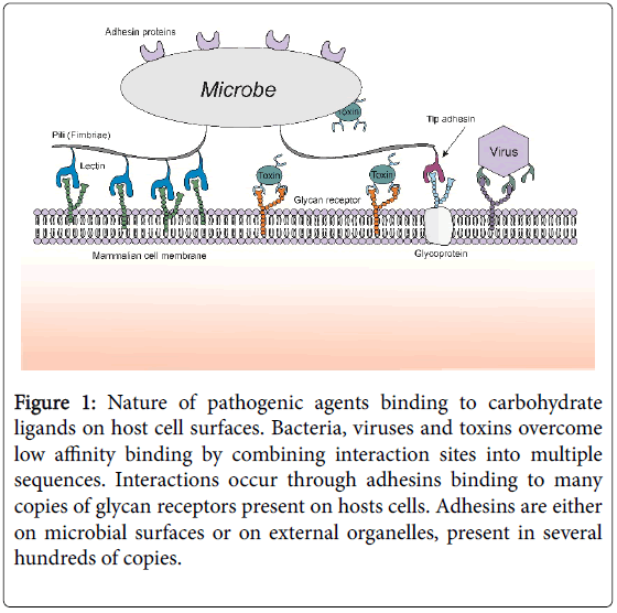
Efficient and stable adhesion is a prerequisite for microbial colonization and invasion of the host cell [5]. The ability of a pathogen to attach to host cells allows it to overcome the natural mechanical shear forces and escape immunological surveillance mechanisms (Figure 1). The initial attachment also enables access to nutrients and gives the pathogen sufficient time to exert its mode of action on the host cell, efficiently deploying their repertoire of virulence factors, including the production of toxins. Pathogens first attach to their host cell using weak non-specific interactions based on physicochemical properties such as charge and hydrophobicity [5]. This initial adsorption is followed by transient interactions, allowing a rolling or gliding motion, with slow movement to sample the host cell surface. This step enables specific adhesin-receptor pairing and allows the targeting of a particular microbe to a specific surface (tissue tropism). The binding pair-wise combinations, on both the guest and host side, can vary in terms of surface molecules which can be a sugar, a protein or a lipid. The most common strategy used by pathogens involves the interaction between lectins (carbohydrate-binding proteins) and host glycoconjugates (glycoproteins, glycolipids and proteoglycans), which are specific for the target tissue [6]. Inversely, microbial surfaces can also be rich in glycoconjugates such as capsules, lipopolysaccharides, and peptidoglycans which bind to host cell lectins [7]. Therefore, research addressing molecules that mediate invasion of a host cell through carbohydrate-lectin mechanisms is a very promising strategy towards inhibiting the initial steps of biothreat agents.
Figure 1: Nature of pathogenic agents binding to carbohydrate ligands on host cell surfaces. Bacteria, viruses and toxins overcome low affinity binding by combining interaction sites into multiple sequences. Interactions occur through adhesins binding to many copies of glycan receptors present on hosts cells. Adhesins are either on microbial surfaces or on external organelles, present in several hundreds of copies.
This review was written with explicit and systematic approach to outline new strategies for preventing and controlling the spread of deadly infectious diseases. Methodology used is based on defined questions, focusing on the role of glycan-based interactions in antiadhesion approaches against biothreat agents. An extensive search of relevant studies by queries in the freely accessible biomedical databases was conducted. Relevant biothreat organisms, with representative examples against bacterial, toxin and viral interactions, were selected and studies from the field of glycobiology, with research work providing significant and promising findings in anti-adhesive therapies, were described. The quality of studies was assessed and the evidence of their impact was summarized.
Carbohydrate-lectin Interactions
The importance of carbohydrates in complex biological processes has been largely underestimated for years. With the analogy to an alphabet in mind, a molecular language of life consisting of nucleic acids and proteins is complemented by glycans [8]. These three biomolecules assure the flow of information from genes to cellular activities and communication between cells at every level of life [9]. Nucleotides and amino acids are translationally predictable due to their linear arrangements, template-based synthesis and fixed linkage structures. In contrast, monosaccharide building blocks can differ in arrangement of atoms in three-dimensional space and they possess the ability to form highly branched molecules. Glycosylation machinery and functions of many glycoconjugates have been described in detail in recent years and specific glycan-lectin pairs were defined as crucial for the interaction of biomolecules during immune recognition, inflammation, and infection of pathogenic organisms [10,11].
The discovery of new lectins dramatically helped to understand the role of glycosylation as a post-translational modification with its extreme diversification emerging from the formation of complex saccharides [9]. Lectins show poor affinity for their monovalent ligands; however, a large body of research has focused on the role of multivalent glycan receptors presented on proteins and lipids which display high avidities for lectins in specific biological processes observed in vivo. Through the mechanisms of evolution, many pathogens are able to express an array of different proteins involved in adhesion and toxicity, with some appearing as multimers [12]. For bacteria, fimbriae or pili are the typical organelles utilised during an initial attachment (Figure 1). In many cases, it is a monomeric lectin with only one binding site at the tip of the fimbriae that is the actual adhesin and the large number of these external structures on the bacterial surface generates multiple anchoring points [13]. Other type of pili can express multiple lectins on one organelle to provide multivalency by a ‘Velcro® mechanism’ [14]. Afimbrial surface adhesins or polysaccharide surface layer can also be involved in the attachment [7]. Additionally, bacteria and viruses constantly change in the heritable traits between successive populations, and due to short generation times, genetic diversity is observed as a large pool of mutants with varying degrees of virulence and glycan-binding affinities. The rapidity, with which some microorganisms mutate, destroyed multiple attempts to control selected diseases by preventative immunizations and pushed the development of other prophylactic and therapeutic approaches [4].
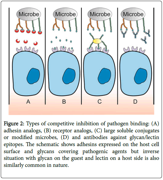
The molecular basis of anti-adhesive strategies comes from the assumption that once the adherence is inhibited or impaired, the host organism is either not infected at all, or it gains significant time advantage for its immune system to overcome a lower load of infectious agents. These treatments might also outcompete and dislocate pathogens that have already attached. The anti-adhesive strategies do not result in the direct death of the pathogenic agent but rather depend on the host’s immune system or physical barrier for clearance. The four general types of competitive inhibition of pathogen binding are adhesin analogs, receptor analogs, large soluble conjugates or modified microbes, and antibodies against glycan/lectin epitopes (Figure 2). The competitive substitutes are either produced synthetically or derived from natural sources, including extracts of milk, fungi, algae, and different parts of plants e.g. from leaves, grains and berries [15,16].
Figure 2: Types of competitive inhibition of pathogen binding: (A) adhesin analogs, (B) receptor analogs, (C) large soluble conjugates or modified microbes, (D) and antibodies against glycan/lectin epitopes. The schematic shows adhesins expressed on the host cell surface and glycans covering pathogenic agents but inverse situation with glycan on the guest and lectin on a host side is also similarly common in nature.
To simplify the description of competitive substitutes in this paragraph, the situation when adhesins are expressed on the host cell surface and glycans are covering pathogenic agent is discussed below (inverse circumstances with glycan on the guest and lectin on a host side is also similarly common in nature). Adhesin analogs (Figure 2A) compete for the attachment with pathogens by blocking their glycan receptors and reducing the number of possible available glycans on pathogenic surface [17]. In this situation, large quantities of foreign protein molecules are required, increasing the chance of immunogenic or toxic effect. Development of appropriate methods to deliver adhesin analogs close to the site of infection is also challenging [18]. Receptor analogs (glycomimetics, adhesin inhibitors) provide overall reduction of available active sites by ‘filling out’ adhesins expressed on host cells (Figure 2B). The use of a cocktail of inhibitors directed towards different carbohydrate specificities has a key advantage of targeting multiple adhesins at the same time [18]. Due to the similarity of the glycans to the structures present on the host cells, they rarely cause immunogenicity or toxicity, even when delivered in excess. Furthermore, glycan receptors can be placed in a large number on different scaffolds (multivalent systems) or expressed by bacterial species derived from natural flora of the body and provided as probiotics (Figure 2C) [19]. Production of antibodies against glycans present on pathogenic agents is a common approach when surface proteins are heavily glycosylated and cover the peptide part which cannot be reached by antibodies (Figure 2D) [18].
Characterization of Carbohydrate-lectin Interactions
Many bioactive oligosaccharides have huge structural complexity in the form of a polysaccharide or as a component of cellular glycoconjugates; however, for a majority of them only a small part of the molecule is participating in receptor binding [7]. Large portions of these molecules serve as scaffolds and act as an orientation tool for the appropriate conformation of the molecule. Hence, it has been generally possible to synthesise ligand structures and design specific glycomimetics, molecules that have structures similar to carbohydrates but result in modified biological properties. The most common approach in the field of glycomimetics is to start from the natural ligand by understanding how the selected protein–glycan interaction occurs and how it can be modified at the molecular level to improve binding properties of alternative molecules.
Lectin and glycan microarrays are two of the key tools used in laboratories to screen molecules and cells for glycans or lectins respectively [20]. Arrays differ in the range of glycans/lectins and the manner in which they are displayed on the chip surfaces as this can be an influencing factor in the recognition of the glycan [21]. In many cases, specific thermodynamically stable conformation is needed for receptor recognition and high binding affinities are only achieved in the presence of specific neighbouring residues. Many of the carbohydrate-lectin interactions can be observed for free molecules in solution but not for tethered forms. Nevertheless, availability of highthroughput screening has simplified a tedious and expensive task of nominating promising ligand candidates previously achieved by testing molecules separately [22].
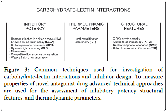
Defining in detail the specific ligand’s primary topology, density and presentation of carbohydrates is of crucial importance and requires several advanced technical approaches (Figure 3). To design alternative structures the priorities will be directed towards a high affinity molecule together with good selectivity to specifically block the defined carbohydrate-lectin binding pair. Structural features, inhibitory potency and thermodynamic parameters are assessed to measure novel antagonist drug potency with a number of basic parameters first determined (Figure 3). The dissociation constant Kd is used routinely as it measures the natural tendency of a complex to separate (dissociate) reversibly into smaller components. However, Kd determination can be technically challenging for multiple binding sites and quite misleading for the potency of the synthetic candidates in vivo . The IC50 parameter is a highly informative measure of how effective a drug or other substance is, and represents the concentration of a drug that is required for 50% inhibition in vitro for a given biological process or its component. IC50 can be easily measured in competition experiments but in general, obtained values are highly dependent on the experimental conditions like the nature of the buffer, flow, and concentrations of bioanalytes, the sensor used but also the type of glycan spacer, its density and architecture. Several powerful techniques (HIA, ELLA, SPR, DLS, microarrays, fluorescence polarisation and weak affinity chromatography), are used to measure inhibitory potency. Isothermal titration calorimetry (ITC) is a labelfree technique providing thermodynamic data especially for glycans in solution, which are highly relevant to in vivo biological function, allowing for further optimization of compounds. Exhaustive description of advantages and challenges of aforementioned techniques were recently reviewed elsewhere [23].
Rational Design of High Affinity Compounds for Antiadhesion Therapies
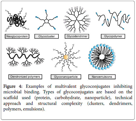
Lectin-carbohydrate interactions are well known to appear in vivo as multivalent systems. The natural monovalent glycan ligands are rarely used for anti-adhesion therapeutics as small single molecules because their interactions would be too weak (in the μM to mM range) [24]. Synthetic glycomimics and glycoconjugates generally would aim at achieving improved topology and binding properties in comparison to the natural ligands [23]. The polymer chemistry field took advantage of the known “glycocluster effect” to enhance lectin avidity by developing a range of linear and spherical multivalent glycoconjugates. Lectin-based design is still the most widely adopted approach for designing a multivalent ligand. It is based on the multimeric lectin structures and attempts to design a multiglycosylated structure that would best correlate with the lectin topology. Selection of a scaffold can significantly improve activity (modification of avidity and specificity) and it is a crucial element during the design of multivalent ligands. Based on structural complexity, technical approach and the backbone selected, several types of multivalent glycoconjugates can be defined (Figure 4).
The synthesis of neoglycoconjugates (NGCs), mostly by random chemical glycation of proteins at reactive residues, has reached considerable levels of availability and sophistication, with various structures, valencies, and conformations available commercially (Figure 4).
Classical multivalent NGCs and glycoproteins have been widely used in array format to screen for and study a great number of carbohydrate-lectin interactions with limited amount of samples needed. Glycoclusters require the synthetically controlled conjugation of a limited number of biologically relevant carbohydrate epitopes, functionalized with linker arms to a multivalent core (chiral carbohydrate scaffold) to combine the advantages of homogeneous small molecular inhibitors with increased valencies. In glycodendrimers an additional branching point is incorporated into the structure leading to multiple layers in globular molecules which contain a central core and much higher valencies due to numerous functionalized end groups. Glycopolymers are synthetic carbohydratecontaining macromolecules, developed either by polymerization of monomers containing carbohydrate moieties or by chemical modifications of functional polymers with carbohydrates using controlled polymerization techniques and click reactions. However, it is still challenging to control precision and reproducibility of glycopolymers to assure higher valencies with different chain lengths, monomer sequence, chain folding, and tertiary structures. Different approaches have resulted in the development of several major types of glyconanoparticles: glycosylated macromolecular structures with metallic, magnetic, semiconductor (quantum dots) and self-assembled cores [25]. Glyconanoparticles combine the multivalent presentation of carbohydrates (glycoclusters) with the special chemiphysical properties. Many of these materials are still in their infancy and the toxicity of their polymer backbone needs to be verified for their approval as therapeutics. However, glyconanoparticles provide much larger valencies, typically in range of 50-150 residues, and they represent the promising trend in glycan-based therapeutics with a rapid development in recent years.
Specific Anti-adhesion Molecules Targeting Bacteria
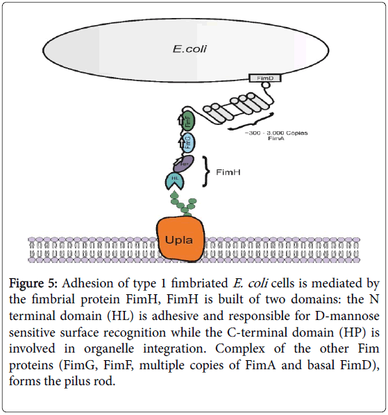
Uropathogenic strains of E. coli (UPEC) are the causative agent of 80 to 90% of urinary tract infections (UTIs). In developed countries more than 80% of UTIs are uncomplicated; however, recurrent infections are a main source of morbidity and health-care cost in this population with further risk of development of resistant strains in case of repeatable antibiotic therapies. UPEC tropism for the bladder tissue is largely mediated by type 1 (Fim) pili (Figure 5) [26]. The rod of the Fim pilus is composed of up to a thousand FimA protein subunits, which are organised in a helical manner to create a force-sensitive cylinder [27]. The distal tip is composed of two adaptor proteins, FimF and FimG, and the two-domain tip adhesin FimH (pilin domain FimHP and lectin domain FimHL). Specifically, the FimHL external domain is a mannose-binding lectin, interacting with mannosylated uroplakin (upla) receptors on the uroepithelium of host cells. FimH binding site can accommodate only one α-mannoside, and once bound using multiple pili, E. coli cells are internalized in an active process that is similar to phagocytosis.
Figure 5: Adhesion of type 1 fimbriated E. coli cells is mediated by the fimbrial protein FimH, FimH is built of two domains: the N terminal domain (HL) is adhesive and responsible for D-mannose sensitive surface recognition while the C-terminal domain (HP) is involved in organelle integration. Complex of the other Fim proteins (FimG, FimF, multiple copies of FimA and basal FimD), forms the pilus rod.
The design of carbohydrate-based inhibitors of FimH-mediated adhesion has attracted a lot of interest both for single molecules and multivalent systems. Various aromatic α-mannosides and oligomannosides with potency in the low nanomolar range were identified and only a few are mentioned below as examples. FimH can reach a 100-fold increase once binding to glycans exposing terminal Manα-(1,3) Manβ(1,4)GlcNAc trisaccharide or oligomannosides [13]. The addition of alkyl chains at the anomeric position of a mannose residue in α-configuration resulted in high affinity ligands. The aryl chain stability is due to the hydrophobic interactions with the “tyrosine gate” residues (Tyr48 and Tyr137) at the binding site.
Exciting examples of multivalent glycoconjugates inhibiting type 1 fimbriae-mediated bacterial adhesion include Octopus glycosides, tetravalent glycocluster based on azamacrocycles, glycodendrimers and hexavalent thiourea-based glycocluster, bifunctional ligands and glyconanodiamonds [28-31]. Adhesion on multivalent glycomaterials and glyconanodiamonds decorated with mannose can be utilized for aggregating E. coli and removing them from polluted water [13].
Anti-adhesion Molecules Targeting Toxin Binding
Shiga toxin
Shiga toxin (Stx) from Shigella dysenteriae type 1, is the prototype toxin for the family of Shiga toxins [32]. When Shiga toxin-producing E. coli (STEC) strains were associated with haemolytic uraemic syndrome (HUS), the scientific community realised that the causative agents were either identical to Shigella -derived or highly related toxins. Two main types of toxin were defined: Shiga toxin type 1 (Stx1), with very high similarity to the original Stx produced by S. dysenteriae type 1, and the immunologically distinct type (Stx2). Epidemiological studies revealed that the variants of Stx range in their ability to cause human diseases and when differing from the parent strains in a few amino acids they can bind to different glycans.
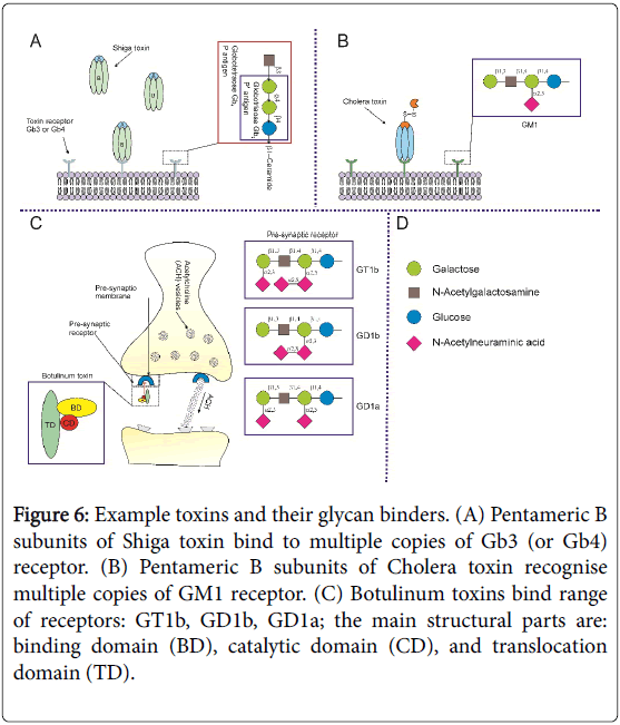
Shiga toxin is composed of two types of subunits, A and B, and has an AB5 molecular configuration as revealed by X-ray crystallography [33]. The monomeric A subunit with a molecular mass of 32 kDa is enzymatically active and non-covalently associated with a homopentamer of B fragments, each with a molecular mass of 7.7 kDa (Figure 6A). The B5 element binds to the cellular toxin receptor, the glycosphingolipid, globotriaosylceramide (Gb3), also known as the Pk trisaccharide. The toxin is then transported from the plasma membrane through early endosomes and the Golgi complex to the endoplasmic reticulum [34]. The A subunit inactivates the ribosome, halting protein synthesis, cleaving a single adenine residue from the 28S ribosomal RNA molecule. Thus to exert toxic catalytic activity in the cytosol of target cells Stx depends on successful binding to Gb3 receptor. Interestingly, Stx2 variant uses Gb4 as a receptor instead of Gb3 and is toxic to pigs, but not to humans.
Figure 6: Example toxins and their glycan binders. (A) Pentameric B subunits of Shiga toxin bind to multiple copies of Gb3 (or Gb4) receptor. (B) Pentameric B subunits of Cholera toxin recognise multiple copies of GM1 receptor. (C) Botulinum toxins bind range of receptors: GT1b, GD1b, GD1a; the main structural parts are: binding domain (BD), catalytic domain (CD), and translocation domain (TD).
One of the first therapeutically available glycodendrimers was the design of STARFISH [35]. It is composed of an oligovalent dendron with Gb3 trisaccharide analogs (Pk) attached to a glucose pentavalent core with inhibitory activity against Stx improved 1 to 10 million fold. The STARFISH inhibitor was further modified to create (S)-PolyBAIT glycopolymers, composed of a monomer containing the Pk glycan linked to cyclic pyruvate (CP) ketal [36]. CP binds serum amyloid P component (SAP), an endogenous protein that is able to target bound ligands for clearance of Stx. Mice subcutaneously injected with a lethal dose of Stx1 were fully protected against the toxin. Interestingly, in a similar experiment when Pk and CP were randomly distributed in glycopolymer, the mice developed severe signs of toxin effect proving that not only the ligand but also the backbone and distribution must be carefully designed for deactivation of toxins [36].
Cholera toxin
Cholera toxin (Ctx) is secreted by Vibrio cholerae and is responsible for the acute diarrhoeatic symptoms normally associated with cholera infection. The effects of Ctx in humans can be drastic resulting in very rapid fluid loss from the intestine, causing severe dehydration. It belongs to the AB5 family of toxins, similar to Stx, and contains the five identical B subunits bound to one catalytic A subunit (A1 and A2 parts connected by disulphide bridge). During the first stage of toxin action, the pentameric ring of B subunits of the Ctx binds to gangliosides containing pentasaccharide, GM1, (monosialotetrahexosylganglioside) found in lipid rafts on the target cells (Figure 6B). Dendrimers with GM1 and pentavalent inhibitors were generated to inhibit the Ctx cellular toxicity [37,38]. In the screening of 35 galactosides the mnitrophenyl α-D-galactopyranoside was nominated as an inhibitor of promising potency with a sub-millimolar IC50 value and bivalent ligands with a longer aglycon incorporating a piperazine motif were demonstrated to be an effective way of blocking Ctx [39,40]. In a screen of a small library of nonhydrolyzable mimics of GM1 ganglioside a submillimolar ligand was recommended for the design of multivalent glycoconjugates [41]. The synthesis of a highly potent, branched pentavalent ligand with appropriate ring size and linker arm lengths was achieved with well-defined geometry and shown to inhibit cholera toxin [42].
Botulinum neurotoxin
Botulinum neurotoxin (BoNT) induces a potentially fatal paralytic condition known as “botulism”. It is currently the most potent toxin known (LD50 of 1–5 ng/kg weight), existing in 7 toxin types (BoNT/A through to BoNT/G) that are further distinguished by antibodies into distinct serotypes. BoNTs are produced by the gram-positive, sporeforming, anaerobic bacterium, Clostridum botulinum. Structural studies revealed that the BoNTs share the same domain organization and are expressed as a single inactive 150 kDa polypeptide chains [43]. Tissue proteinases cleave the peptide, connected by a single disulfide bond, into a heavy chain (H) of 100 kDa and a light chain (L) of 50 kDa (Figure 6C). The H chain then binds to its specific presynaptical membrane receptor of the nerve muscle junctions, typically a polysialoganglioside, GT1b, GD1b and GD1a (an oligosaccharide in which one or more sialic acid molecules are linked to the carbohydrate moiety) to induce membrane translocation and endocytosis by intracellular synaptic vesicles [44]. The L chain cleaves members of the soluble N-ethylmaleimide-sensitive-factor attachment receptor (SNARE) family, leading to the inhibition of synaptic vesicular fusion exocytosis, blocking the release of the neurotransmitter acetylcholine at the neuromuscular junction, consequently stopping neural transmission resulting in long-lasting flaccid paralysis of muscles. Hence, BoNTs uses an interaction mode that requires at least two different receptors, oligosaccharide portion of a polysialoganglioside and a protein receptor.
To neutralize the circulating BoNTs the currently used antidotes are equine antitoxin antibodies for treatment of adult patients and human antitoxin is recommended for infants (IgG preparation from the blood of volunteers vaccinated with pentavalent botulinum toxoid). Many studies attempted for designing small molecules, peptides and aptamers or testing natural substances for inhibitors of botulinum activity. The SELEX approach was utilised to generate high affinity ssDNA aptamers against BoNTs [45]. BoNT binding to the ganglioside receptor can be inhibited by quinic acid (a cyclic polyol found in coffee beans) at a concentration of 10 mM [46]. Lectins, from Limax favus and Triticum vulgaris showed significant inhibition of various BoNTs as competitive antagonists [46]. Thearubigin, extracted from black tea, binds with the BoNT in a dose-dependent manner inhibiting muscle paralysis in mouse models [47]. Like antibody based therapy, the treatment window for such agents is short, since they can only target the antigen at the circulation level. Thus, further studies leading to high availability and easy access of to the BoNT binders are critically needed.
DC-SIGN Inhibitors as Viral Anti-adhesives
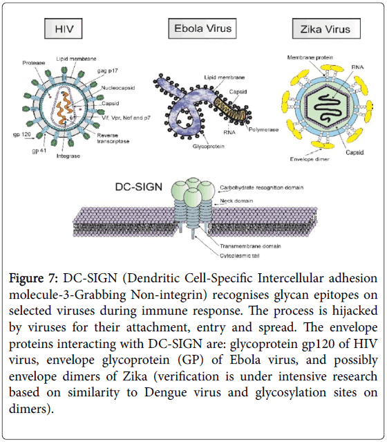
The immune system can also use host lectins to identify and bind glycans displayed on pathogenic surfaces; however, in some cases the microorganisms can reroute this process and use it for invasion. DCSIGN (Dendritic Cell-Specific Intercellular adhesion molecule-3- Grabbing Non-integrin) is a C-type lectin receptor present on the surface of antigen presenting cells (dendritic cells) and macrophages subpopulations (Figure 7). DC-SIGN recognises and binds through multivalent glycan–protein interactions to surface-rich mannosecontaining glycans, (Man)9(GlcNAc)2, a branched oligosaccharide that is commonly found in multiple copies on pathogen glycoproteins including several viruses, bacteria and fungi [23]. DC-SIGN is also able to recognize branched fucosylated structures with terminal galactose residues, such as the Lewis antigens. This binding interaction on macrophages activates phagocytosis [48].
Figure 7: DC-SIGN (Dendritic Cell-Specific Intercellular adhesion molecule-3-Grabbing Non-integrin) recognises glycan epitopes on selected viruses during immune response. The process is hijacked by viruses for their attachment, entry and spread. The envelope proteins interacting with DC-SIGN are: glycoprotein gp120 of HIV virus, envelope glycoprotein (GP) of Ebola virus, and possibly envelope dimers of Zika (verification is under intensive research based on similarity to Dengue virus and glycosylation sites on dimers).
Human Immunodeficiency Virus
More than a decade ago, human immunodeficiency virus (HIV-1) was shown to target DC-SIGN using its envelope glycoprotein gp120 [49]. However, it was able to escape degradation in lytic compartments using DCs as a Trojan horse to invade the host organism. Thus for some pathogens, including viruses like HIV, Ebola, or Dengue, this recognition event, rather than protecting the host, helps in infection by promoting viral replication and transmission.
The HIV envelope protein, gp120, remains effectively hidden from antibodies because its surface is shielded by glycans. Thus, isolation of antibodies that bind the high mannose moieties of the gp120 was one of the biggest breakthroughs in the glycobiology field [50,51]. The 2G12 clone is the best described broadly neutralizing antibody, which slows the entry of the virus into cells and inhibits replication [52]. The PG9 clone binds to the glycans from both gp120 and gp140 envelope proteins. Glycodendrimers containing mannose residues were shown to inhibit binding of the gp120 mannosylated HIV envelope protein [53]. A recombinant dimeric DC-SIGN was blocked by oligomannose dendron with IC50 values in the nanomolar range [53]. Moreover, nanoparticles bearing mannose dendrimer mimics and a high mannose dendrimer were used for blocking DC-SIGN mediated HIV infection in cellular and human uterine cervix explant models [54,55]. The incorporation of 1,2-mannobioside epitopes into gold glyconanoparticles (Au-GNPs) with valencies up to 50 or 60 copies completely inhibited the interaction of DC-SIGN with gp120 [54].
Ebola Virus
Ebola haemorrhagic fever is a rare and deadly disease caused by infection with one of the Ebola virus species of the Filoviridae family of negative-stranded RNA viruses. It was first discovered in 1976 near the Ebola River in Congo, later found in several African countries. Ebola disease is a rather local public health threat in Africa with sporadic outbreaks affecting others worldwide due to imported infections. However, mortality rate is extremely high, ranging from 30% to 90%, depending on the virus type and estimation methodologies. The WHO reported cases in the West African countries during the recent 2013-2016 outbreak were in a total of 28,657 suspected cases and 11,325 deaths, yielding 40% mortality rate [55,56]. Ebola virus entry into target cells is mediated by a single viral surface glycoprotein GP [57]. Similar to gp120 of HIV, the Ebola’s GP is heavily glycosylated and interacts with several cellular receptors, including C-type lectins DC-SIGN and L-SIGN [58]. During early steps of virus infection GP attachment stimulates virus macropinocytosis [59].
The glycopolymer field has addressed Ebola inhibition and constructed multi-block glycopolymers with acrylate monomers consisting of mannose, glucose, and fucose residues [60]. The resulting “glycodendronanoparticles” are the most highly branched glycodendrimeric constructs with diameters of up to 32 nm corresponding to 1,620 glycan units. The dendrimers with certain clustered mannose structures were capable of blocking viral binding to DC-SIGN (at picomolar concentrations) in a model where Tlymphocytes and human dendritic cells were infected by the Ebola virus [58]. Moreover, soluble glycofullerenes have shown interesting antiviral activity in an Ebola pseudotyped infection model, thus opening new perspectives for their applications [61]. Serum-derived circulating mannose-binding lectins (MBL) and recombinant human MBL (rhMBL) demonstrated the potential utility against Ebola [62]. Griffithsin, is a red algal lectin, first reported as a novel anti-HIV protein [62], having an average EC50 of 40 pM. The glycoprotein (GP) on the surface of the Ebola virus has similar N-linked mannose-rich glycans as on the HIV virus, allowing griffithsin to bind GP of the Ebola virus in a similar manner. Thus, griffithsin which possesses good stability over a satisfactory pH and temperature range, can be used as broad spectrum antiviral therapeutic against Ebola infection [63,64]. Smaller griffithsin-derived peptides were also reported to have glycanbinding and HIV-inhibitory properties but have not been evaluated yet as therapeutics against Ebola [65].
Zika Virus
In 2016, WHO declared international public health emergency over Zika virus which is spreading explosively across Central and South America. The virus is named after the Zika forest in Uganda where it was discovered in 1947 and was thought for decades to be practically harmless. However, it turned out to be responsible for neurological abnormalities in developing fetuses and serious birth defects observed as a spike in cases of micro-encephalitis in Brasil in 2015 [66]. It belongs to a family of Flaviviruses (which also includes the Dengue, yellow fever and West Nile viruses), a single stranded, positive-sense RNA virus with a 10.7 Kb genome encoding a single polyprotein that is cleaved into three structural proteins (C, precursor M/M, and E) and seven non-structural [67]. The envelope of Flaviviruses , determined by cryo-electron microscopy, was shown to be made up of 180 copies of two different proteins, the envelope (E) protein and membrane (M) protein, in which 90 E dimers completely cover the viral surface [68,69]. E protein is a glycoprotein responsible for virus entry, representing a major target of neutralizing antibodies for Flaviviruses , including Zika virus, Dengue virus, and other mosquito-transmitted viruses from the same family, was shown to bind to DC-SIGN via the glycans on E proteins of the mature virion [70]. It has been already demonstrated that broadly neutralizing antibodies for E-dimer epitope isolated from patients with Dengue disease neutralize Zika as potently as they neutralize Dengue virus [71]
E protein of Zika virus differs from the other known Flaviviruses in the ~10 amino acids that surround the Asn154 glycosylation site in the virus shell [68]. The virus projects a glycosylation site outward and glycans are attached to the viral protein surface at this site. Thus, differences in carbohydrate molecules in this region may influence virus transmission and explain its tissue tropism or variable disease progression in different parts of the world [72]. Furthermore, glycosylation is a promising investigation target in context of potential receptors for anti-adhesive therapies. Zika is spread by mosquitos and the primary defense against it is to remove breeding sites and avoid being bitten. Other protective approaches are desperately needed to decrease the chance of infection and to protect against an inflammation of the fetal brain, contracted in the first months of pregnancy.
Influenza Glycobiology Success Stories
Epidemic infections with influenza virus continue to occur globally with an annual attack rate estimated at 5%-10% in adults. Illnesses is usually mild but can be a serious health concern causing significant morbidity and mortality among high-risk groups (the very young, elderly or chronically ill), even in nations with the most advanced healthcare systems. The life cycle of the influenza virus involves the binding of multiple viral hemagglutinin (HA) molecules to α(2-3)- linked sialic acid (avian receptor) or α(2-6)-linked sialic acid (human receptor) on host cell surfaces of epithelium in respiratory tract [8]. Neuraminidase (NA) is the receptor-destroying enzyme which ensures newly made virus release and maturation by cleaving the same glycans [73]. Each influenza particle is built of approximately 500 copies of HA trimers and 100 copies of NA tetramers on its surface.
The most successful glycobiology drugs approved for treatment and chemoprophylaxis of influenza, and still active against the majority of recently circulating subtypes of influenza, are the antiviral compounds zanamivir (Relenza) and oseltamivir (Tamiflu) [74,75]. Both antiviral drugs mimic the natural sialic acid substrate for the virus NA enzyme; however, they bind much tighter, with nanomolar affinity, leading to the blocking of viral budding and reducing viral spread into other cells [73]. Oral oseltamivir and inhaled zanamivir contribute to reducing mortality and the duration of influenza. Unfortunately, the development of resistance to antiviral agents, like the commonly used neuraminidase inhibitors, was reported [76]. Alternative inhibitors, overcoming acquired resistance to oseltamivir, were searched in recent years and numerous successes were reported that target the influenza virus. Peramivir, with efficacy similar to that of oseltamivir, has recently been approved in the USA by the FDA to treat acute uncomplicated influenza in adults and be given as intravenous injections Laninamivir octanoate, other example of NA inhibitors was shown to be effective if given as a single inhalation [77,78]. Laninamivir has been approved for influenza treatment and for prophylaxis in Japan and is under clinical evaluations in other countries. In conclusion, the dynamic field of glycobiology has integrated very well into the never-ending battle between microbes and drug developers.
Summary
The continuous emergence of antibiotic resistant pathogens is a clear indication that alternative approaches are needed in the fight against infectious diseases. The use of anti-adhesion molecules is a novel approach that is not only effective in preventing infection but also prevents the susceptibility of deadly pathogens becoming resistant to antibiotics. The principle of using anti-adhesion molecules has already been shown to be effective in the treatment of influenza; therefore, translating the same principle to other biothreat agents that mediate invasion of a host cell through carbohydrate-lectin mechanisms is a very promising strategy. Understanding the surface molecules displayed on biothreat agents such as pathogenic bacteria, toxins and viruses, is essential to further the development of antiadhesion based therapies and preventing the spread of antibiotic resistant pathogens.
Acknowledgements
The authors thank the following agencies for financial support: European Defence Agency (EDA) through the DCLAW (Decontamination by Carbohydrate Lectin Affinity Wipes) project (A-1152-RT-GP) and the European Union FP7 programme in support of the NAPES (Next Generation Analytical Platforms for Environmental Sensing) project (Project Number 604241).
References
- Tanwar J, Das S, Fatima Z, Hameed S (2014) Multidrug resistance: an emerging crisis. Interdiscip Perspect Infect Dis 2014: 541340.
- Alekshun MN1, Levy SB (2007) Molecular mechanisms of antibacterial multidrug resistance. Cell 128: 1037-1050.
- Barlow M1 (2009) What antimicrobial resistance has taught us about horizontal gene transfer. Mol Biol 532: 397-411.
- Wang Q, Ling C (2014) Addressing the global need to combat multidrug resistance: carbohydrates may hold the key. Future Med Chem 6: 1539-1543.
- Dufrêne YF1 (2015) Sticky microbes: forces in microbial cell adhesion. Trends Microbiol 23: 376-382.
- Singh RS, Walia AK (2014) Microbial lectins and their prospective mitogenic potential. Crit Rev Microbiol 7828: 329-347.
- Tan FYY, Tang CM, Exley RM (2015) Sugar coating: Bacterial protein glycosylation and host–microbe interactions. Trends Biochem Sci 40: 342-350.
- Weiss AA, Iyer SS (2007) Glycomics Aims To Interpret the Third Molecular Language of Cells. Microbe 2: 489-497.
- Gabius HJ1, Siebert HC, André S, Jiménez-Barbero J, Rüdiger H (2004) Chemical biology of the sugar code. Chembiochem 5: 740-764.
- Gamblin DP1, Scanlan EM, Davis BG (2009) Glycoprotein synthesis: an update. Chem Rev 109: 131-163.
- Spiro RG (2002) Protein glycosylation: nature, distribution, enzymatic formation, and disease implications of glycopeptide bonds. Glycobiology 12: 43R-56R.
- Gabius HJ1, Manning JC2, Kopitz J3, André S2, Kaltner H2 (2016) Sweet complementarity: the functional pairing of glycans with lectins. Cell Mol Life Sci 73: 1989-2016.
- Hartmann M, Lindhorst TK (2011) The Bacterial Lectin FimH, a Target for Drug Discovery–Carbohydrate Inhibitors of Type 1 Fimbriae-Mediated Bacterial Adhesion. 2011: 3583-360.
- Chahales P, Thanassi DG (2015) Structure, Function, and Assembly of Adhesive Organelles by Uropathogenic Bacteria. Microbiol Spectr 3: 5.
- Peterson R, Cheah WY, Grinyer J Packer N (2013) Glycoconjugates in human milk: Protecting infants from disease. Glycobiology 23: 1425-1438.
- Signoretto C, Canepari P, Stauder M, Vezzulli L, Pruzzo C (2012) Functional foods and strategies contrasting bacterial adhesion. Curr Opi. Biotechnol 23: 160-167.
- Krachler AM, Orth K (2013) Targeting the bacteria-host interface: strategies in anti-adhesion therapy. Virulence 4: 284-294.
- Cozens D1, Read RC (2012) Anti-adhesion methods as novel therapeutics for bacterial infections. Expert Rev Anti Infect Ther 10: 1457-1468.
- Rasko DA1, Sperandio V (2010) Anti-virulence strategies to combat bacteria-mediated disease. Nat Rev Drug Discov 9: 117-128.
- Katrlík J1, Svitel J, Gemeiner P, Kozár T, Tkac J (2010) Glycan and lectin microarrays for glycomics and medicinal applications. Med Res Rev 30: 394-418.
- Rillahan CD, Paulson JC (2011) Glycan microarrays for decoding the glycome. See comment in PubMed Commons below Annu Rev Biochem 80: 797-823.
- Roy R, Murphy PV, Gabius H (2016) Multivalent Carbohydrate-Lectin Interactions: How Synthetic Chemistry Enables Insights into Nanometric Recognition. Molecules 21: 629.
- Cecioni S1, Imberty A, Vidal S (2015) Glycomimetics versus multivalent glycoconjugates for the design of high affinity lectin ligands. Chem Rev 115: 525-561.
- Reina JJ, Bernardi A (2012) Carbohydrate mimics and lectins: a source of new drugs and therapeutic opportunities. See comment in PubMed Commons below Mini Rev Med Chem 12: 1434-1442.
- Marradi M1, Chiodo F, García I, Penadés S (2013) Glyconanoparticles as multifunctional and multimodal carbohydrate systems. Chem Soc Rev 42: 4728-4745.
- Lillington J1, Geibel S1, Waksman G2 (2014) Biogenesis and adhesion of type 1 and P pili. Biochim Biophys Acta 1840: 2783-2793.
- Choudhury D, Thompson A, Stojanoff V, Langermann S, Pinkner J, et al. (1999) X-ray structure of the FimC-FimH chaperone-adhesin complex from uropathogenic Escherichia coli. Science 285: 1061-1066.
- Dubber M, Sperling O, Lindhorst TK (2006) Oligomannoside mimetics by glycosylation of octopus glycosides and their investigation as inhibitors of type 1 fimbriae-mediated adhesion of Escherichia coli Org. Biomol. Chem 3: 3901-3912.
- König, Burkhard, Fricke T, Waßmann A, Krallmann-Wenzel U, et al. (1998) α-Mannosyl Clusters Scaffolded on Azamacrocycles: Synthesis and Inhibitory Properties in the Adhesion of Type 1 Fimbriated Escherichia co / i to Guinea Pig Erythrocytes. Tetrahedron Lett 39: 2307-2310.
- Lindhorst TK, Kieburg C, Krallmann-wenzel U (1998) Inhibition of the type 1 fimbriae-mediated adhesion of Escherichia coli to erythrocytes by multiantennary @ -mannosyl clusters: The effect of multivalency. Glycoconj J 613.
- Barras A, Fernando Ariel Martin OB, Baumann JS, Ghigo JM, Rabah Boukherroub CB, et al. (2013) Glycan-functionalized diamond nanoparticles as potent E. coli anti-adhesives. Nanoscale 5: 2307-2316.
- Bauwens A, Betz J, Meisen I, Kemper B, Karch H, et al. (2013) Facing glycosphingolipid-Shiga toxin interaction?: dire straits for endothelial cells of the human vasculature. Cell Mol Life Sci 70: 425-457.
- Hagnerelle X, Lambert O, Marco S, Rigaud JL, Johannes L, et al. (2002) Two-dimensional structures of the Shiga toxin B-subunit and of a chimera bound to the glycolipid receptor Gb3. Journal of Structural Biology 139: 113-121.
- Ling H1, Boodhoo A, Hazes B, Cummings MD, Armstrong GD, et al. (1998) Structure of the shiga-like toxin I B-pentamer complexed with an analogue of its receptor Gb3. Biochemistry 37: 1777-1788.
- Kitov PI, Sadowska JM, Mulvey G, Armstrong GD, Ling H, et al. (2000) Shiga-like toxins are neutralized by tailored multivalent carbohydrate ligands. Nature 403: 669-672.
- Kitov PI, Mulvey GL, Griener TP, Lipinski T, Solomon D, et al. (2008) In vivo supramolecular templating enhances the activity of multivalent ligands: A potential therapeutic against the Escherichia coli O157 In vivo supramolecular templating enhances the activity of multivalent ligands?: A potential therapeutic against the Escherichia coli O157 AB��? Toxins. PNAS 105: 16837-16842.
- Pukin AV1, Branderhorst HM, Sisu C, Weijers CA, Gilbert M, et al. (2007) Strong inhibition of cholera toxin by multivalent GM1 derivatives. Chembiochem 8: 1500-1503.
- Branson TR, Mcallister TE, Garcia-hartjes J, Fascione MA, Ross JF, et al. (2014) Multivalent Inhibitors Very Important Paper A Protein-Based Pentavalent Inhibitor of the Cholera Toxin B. Angew Chem Int Ed 1-6.
- Minke WE, Roach C, Hol WG, Verlinde CL (1999) Structure-based exploration of the ganglioside GM1 binding sites of Escherichia coli heat-labile enterotoxin and cholera toxin for the discovery of receptor antagonist. Biochemistry 5684-5692.
- Pickens JC, Mitchell DD, Liu J, Tan X, Zhang Z, et al. (2004) Nonspanning Bivalent Ligands as Improved Surface Receptor Binding Inhibitors of the Cholera Toxin B Pentamer. Chem Biol 11: 1205-1215.
- Cheshev, Pavel, Morelli L, Marchesi M, Podlipnik C, et al. (2010) Synthesis and Affinity Evaluation of a Small Library of Bidentate Cholera. Chem Eur J 16: 1951-1967.
- Liu J, Zhang Z, Tan X, Hol WGJ, Verlinde CLMJ, et al. (2005) Protein Heterodimerization through Ligand-Bridged Multivalent Pre-organization?: Enhancing Ligand Binding toward Both Protein Targets. 127: 2044-2045.
- Swaminathan S, Eswaramoorthy S (2000) Structural analysis of the catalytic and binding sites of Clostridium botulinum neurotoxin B. Nat Struct Biol 7: 693-699.
- Montal M (2010) Botulinum neurotoxin: a marvel of protein design. Annu Rev Biochem 79: 591-617.
- Fan M, McBurnett SR, Andrews CJ, Allman AM, Bruno JG, et al. (2008) Aptamer selection express: a novel method for rapid single-step selection and sensing of aptamers. J Biomol Tech 19: 311-319.
- Cai S, Singh BR (2007) Strategies to design inhibitors of Clostridium botulinum neurotoxins. See comment in PubMed Commons below Infect Disord Drug Targets 7: 47-57.
- Satoh E, Ishii T, Shimizu Y, Sawamura SI, Nishimura M (2002) A mechanism of the thearubigin fraction of black tea (Camellia sinensis) extract protecting against the effect of tetanus toxin. J Toxicol. Sci 27: 441-447.
- van Liempt E1, Bank CM, Mehta P, Garciá-Vallejo JJ, Kawar ZS, et al. (2006) Specificity of DC-SIGN for mannose- and fucose-containing glycans. FEBS Lett 580: 6123-6131.
- Torensma R, Van Vliet SJ, Van Duijnhoven GCF, Middel J, Kewalramani VN, et al. (2000) DC-SIGN , a dendritic cell-specific HIV-1- binding protein that enhances trans-infection of DC-SIGN, a Dendritic Cell-Specific HIV-1-Binding Protein that Enhances trans -Infection of T Cells. Cell 100: 587-597.
- Pejchal R, Doores KJ, Walker LM, Khayat R, Huang P, et al. (2012) A potent and broad neutralizing antibody recognizes and penetrates the HIV glycan shield. Science 334: 1097-1103.
- Walker LM, Huber M, Doores KJ, Falkowska E, Pejchal R, et al. (2011) Broad neutralization coverage of HIV by multiple highly potent antibodies. Nature 477: 466-470.
- Platt EJ, Gomes MM, Kabat D, Piatt EJ (2012) Kinetic mechanism for HIV-1 neutralization by antibody 2G12 entails reversible glycan binding that slows cell entry. PNAS 109: 7829-7834.
- Wang SK, Liang PH, Astronomo RD, Hsu TL, Hsieh SL, et al. (2008) Targeting the carbohydrates on HIV1: Interaction of oligomannose dendrons with human monoclonal antibody 2G12 and DC-SIGN. Proc Natl Acad Sci USA 105: 3690-3695.
- Martínez O, Bedoya LM, Marradi M, Clavel C, Alcamí J (2009) Multivalent Manno-Glyconanoparticles Inhibit DC-SIGN-Mediated HIV-1 Trans-Infection of Human T Cells. ChemBioChem 20: 1806-1809.
- Berzi, Angela, Reina JJ, Ottria R, Sutkeviciute I, et al. (2012) A glycomimetic compound inhibits DC-SIGN- mediated HIV infection in cellular and cervical explant models. AIDS 26: 127-137.
- Spengler JR, Ervin ED, Towner JS, Rollin PE, Nichol ST (2008) Perspectives on West Africa Ebola Virus Disease Outbreak.22: 2013-2016.
- Lin G, Simmons G, Pöhlmann S, Ni H, Leslie GJ, et al. (2003) Differential N-Linked Glycosylation of Human Immunodeficiency Virus and Ebola Virus Envelope Glycoproteins Modulates Interactions with DC-SIGN and DC-SIGNR. J Virol 77: 1337.
- Alvarez CP, Lasala F, Carrillo J, Muñiz O, Corbí AL, et al. (2002) C-type lectins DC-SIGN and L-SIGN mediate cellular entry by Ebola virus in cis and in trans. J Virol 76: 6841-6844.
- Nanbo A, Imai M, Watanabe S, Noda T, Takahashi K, et al. (2010) Ebolavirus Is Internalized into Host Cells via Macropinocytosis in a Viral Glycoprotein-Dependent Manner. PLoS Pathog 6: 9.
- Choi JH, Croyle MA (2013) Emerging targets and novel approaches to Ebola virus prophylaxis and treatment. BioDrugs 27: 565-583.
- Luczkowiak J, Mun~oz A, Macarena Sa´nchez-Navarro RRV, Ginieis A, Illescas BM, et al. (2013) Glycofullerenes Inhibit Viral Infection. Biomacromolecules 11: 431-437.
- Michelow IC, Lear C, Scully C, Prugar LI, Longley CB, et al. (2011) High-dose mannose-binding lectin therapy for Ebola virus infection. J Infect Dis 203: 175-179.
- Mori T, Keefe BRO, Ii CS, Bringans S, Gardella R, et al. (2005) Isolation and Characterization of Griffithsin, a Novel HIV-inactivating Protein, from the Red Alga Griffithsia sp. J Biol Chem 280: 9345-9353.
- Kaur R, Mehan S, Kalra S (2014) Ebola Virus: Current Therapeutic Approaches. Int J Recent Adv Pharm Res 4: 1-4.
- Micewicz ED, Cole AL, Jung C, Luong H, Phillips ML, et al. (2010) Grifonin-1: A Small HIV-1 Entry Inhibitor Derived from the Algal Lectin , Griffithsin. PLoS ONE 5: 1-11.
- Zanluca C, Melo VC, Mosimann AL, Santos GI, Santos CN, et al. (2015) First report of autochthonous transmission of Zika virus in Brazil. Mem Inst Oswaldo Cruz 110: 569-572.
- Lindenbach BD, Rice CM (2003) Molecular biology of flaviviruses. Adv Virus Res 59: 23-61.
- Sirohi D, Chen Z, Sun L, Klose T, Pierson TC, et al. (2016) The 3.8 Å resolution cryo-EM structure of Zika virus. Science 352: 467-470.
- Kuhn RJ, Zhang W, Rossmann MG, Pletnev SV, Corver J, et al. (2002) Structure of Dengue Virus: Implications for Flavivirus Organization , Maturation , and Fusion. Cell: 108: 717-725.
- Pokidysheva E, Zhang Y, Battisti AJ, Bator-kelly CM, Chipman PR, et al. (2006) Cryo-EM Reconstruction of Dengue Virus in Complex with the Carbohydrate Recognition Domain of DC-SIGN. Cell 124: 485-493.
- Barba-spaeth G, Dejnirattisai W, Rouvinski A, Vaney M, Medits I, et al. (2016) Structural basis of potent Zika-dengue virus antibody cross-neutralization. Nature 536: 48-53.
- Zhu Z, Chan JF, Tee K, Choi GK, Lau SK, et al. (2016) Comparative genomic analysis of pre-epidemic and epidemic Zika virus strains for virological factors potentially associated with the rapidly expanding epidemic. Emerg Microbes Infect 5: e22-11.
- Matrosovich MN, Matrosovich TY, Gray T, Roberts NA, Klenk H (2004) Neuraminidase Is Important for the Initiation of Influenza Virus Infection in Human Airway Epithelium. J Virol 78: 12665-12667.
- von Itzstein M, Wu WY, Kok GB, Pegg MS, Dyason JC, et al. (1993) Rational design of potent sialidase-based inhibitors of influenza virus replication. Nature 363: 418-423.
- Asano N (2003) Glycosidase inhibitors: update and perspectives on practical use. Glycobiology 13: 93R-104R.
- Samson M, Pizzorno A, Abed Y, Boivin G (2013) Influenza virus resistance to neuraminidase inhibitors. Antiviral Res 98: 174-185.
- Mclaughlin MM, Skoglund EW, Ison MG (2016) Peramivir: an intravenous neuraminidase inhibitor. Expert Opin Pharmacother 16: 1889-900
- Sugaya N, Yasuo O (2010) Long-Acting Neuraminidase Inhibitor Laninamivir Octanoate (CS-8958) versus Oseltamivir as Treatment for Children with Influenza Virus Infection. Antimicrob Agents Chemother 54: 2575-2582.
Relevant Topics
- Anthrax Bioterrorism
- Bio surveilliance
- Biodefense
- Biohazards
- Biological Preparedness
- Biological Warfare
- Biological weapons
- Biorisk
- Bioterrorism
- Bioterrorism Agents
- Biothreat Agents
- Disease surveillance
- Emerging infectious disease
- Epidemiology of Breast Cancer
- Information Security
- Mass Prophylaxis
- Nuclear Terrorism
- Probabilistic risk assessment
- United States biological defense program
- Vaccines
Recommended Journals
Article Tools
Article Usage
- Total views: 14714
- [From(publication date):
September-2016 - Jul 18, 2025] - Breakdown by view type
- HTML page views : 13606
- PDF downloads : 1108
