Case Report Open Access
Coronectomy (Intentional Partial Odontectomy) in Lower Third Molar as an Alternative Treatment: Report of Two Cases
Eduardo D1*, Julierme FR1, Ana Paula SC1, Fan Song MD3, Celso KS1 and José WN21Dental School of Araçatuba, Universidade Estadual Paulista (UNESP), São Paulo, Brazil
2Faculty of Dentistry, Universidade Federal de Campina Grande (UFCG), Paraíba, Brazil
3The Second Affiliated Hospital, Sun Yat-Sen University, Guangzhou, China
- *Corresponding Author:
- Eduardo Dias-Ribeiro
DDS, MSc, Department of Oral and Maxillofacial Surgery
Dental School of Araçatuba, Universidade Estadual Paulista
(UNESP) Araçatuba 16015050, Brazil
Tel: +55-83-99031968
E-mail: eduardodonto@yahoo.com.br
Received Date: December 15, 2014; Accepted Date: March 10, 2015; Published Date: March 17, 2015
Citation: Eduardo D, Julierme FR, Ana Paula SC, Fan Song MD, Celso KS, et al. (2015) Coronectomy (Intentional Partial Odontectomy) in Lower Third Molar as an Alternative Treatment: Report of Two Cases. J Oral Hyg Health 3:173. doi: 10.4172/2332-0702.1000173
Copyright: © 2015 Eduardo D, et al. This is an open-access article distributed under the terms of the Creative Commons Attribution License, which permits unrestricted use, distribution, and reproduction in any medium, provided the original author and source are credited.
Visit for more related articles at Journal of Oral Hygiene & Health
Abstract
The principle of coronectomy or intentional partial odontectomy is the removal of the tooth crown, leaving the root in situ. This technique aims to prevent damage to inferior alveolar nerve while applying to removal a third molar or posterior tooth impacted in mandible. In this study, we report two clinical cases with the impacted lower third molar presented roots in close proximity to the mandibular canal and by intentional partial odontectomy. Neurosensory deficits, postoperative infection, periods off follow up and surgical outcomes were emphasized in this study. We concluded that the intentional partial odontectomy is a foreseeable technique and easy to perform in an outpatient setting. It is an alternative procedure in the extraction of impacted lower third molar that has a close relationship with mandibular canal.
Keywords
Tomography; Odontectomy; Mandibular nerve; Third molar; Impacted tooth
Introduction
Coronectomy or intentional partial odontectomy is the removal of the crown of the tooth, leaving the root in situ. This technique, when applied to the removal of a third molar or included posterior tooth in the jaw, is intended to prevent damage to the inferior alveolar nerve [1-7].
In the 1970s, clinical, radiographic and histological experimental studies evaluated the dental roots could submerge within the soft tissues which was also known as "burial root" at the time, it was believed that the maintenance of these roots in their alveoli preserved the height of the alveolar ridge, and consequently it could improve the adaptability and stability of conventional prostheses [8-10].
However, from the 1990s, the emphasis has been changed and some studies evaluated surgery for removal of the mandibular third molar, the close relationship of their roots and the mandibular canal and the risk factors for injury in the inferior alveolar nerve [1-7,11-24].
Currently, the panoramic radiograph suggests a close relationship between the inferior alveolar nerve and the roots of the third molar while the cone beam computed tomography (CBCT) could further predict the possible nerve injury that taken by a performance of removal. Where an intimate relationship is confirmed, the technique of intentional partial odontectomy may be indicated [12,14,24].
This study reported two clinical cases, with impacted lower third molar whose roots in an intimate relationship with the mandibular canal, were treated by intentional coronectomies. Postoperative outcomes were evaluated, including neurosensory deficit, postoperative infection and effectiveness of surgical technique.
Case report
Patients and Interventions:
To standardize the treatment, all reported clinical cases were performed using the same surgical technique and all procedures were performed by the same surgeon. All patients in this study sought care for the extraction of third molars in Oral Surgery Service in Dental Center of Studies and Research (COESP), João Pessoa, Paraíba, Brazil. Patients were asked to sign the consent form and informed to such guidelines and rules that regulated research involving human subjects, adopted by National Health Council.
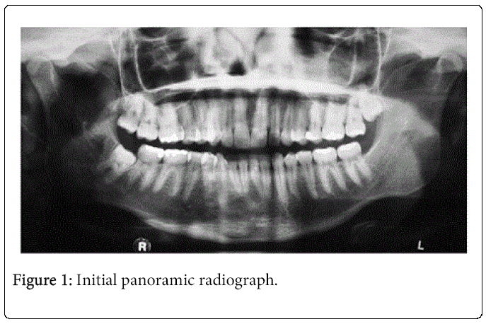
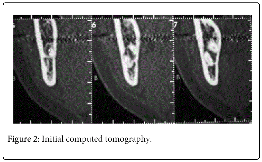
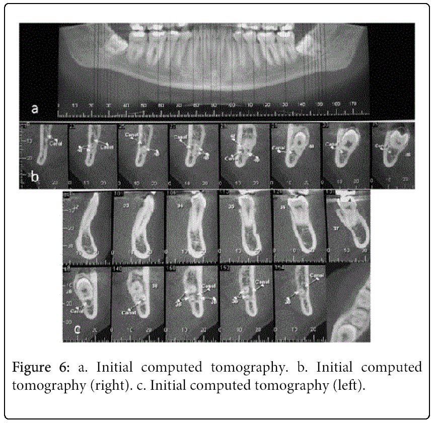
Medical history and physical examination were performed and there was found no change in local or systemic disorders while both patients were evaluated to endure the completion of the surgical procedure. Intraoral examination revealed the lower third molars of both patients presented partial impaction and the crowns were somehow below the mucosa. Therefore, it was requested panoramic radiograph where it was possible to visualize the location of third molars, consequently, both molars were indicated to removal. However, the cases presenting in the study had lower third molars with roots close with the inferior alveolar nerve (Case 1: periapical radiolucent area and darkening of roots. Case 2: darkening of roots and curving of root). Therefore, it was requested CBCT and parasagittal sections was possible to verify the presence of the inferior alveolar nerve and its relationship with the roots of the mandibular third molar.
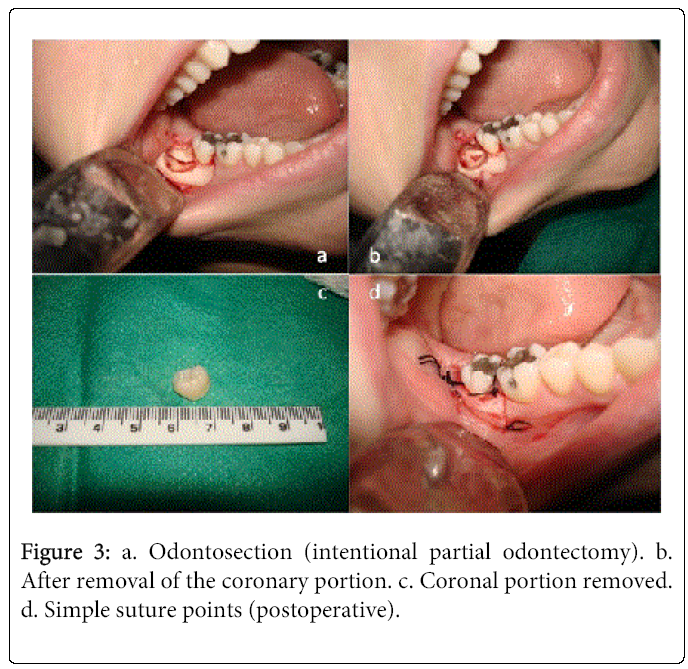
After the diagnosis, surgical treatment was followed basing on the principle of the technique of intentional partial odontectomy. All procedures were performed under local anesthesia that mepivacaine hydrochloride 2% with epinephrine 1:100.000 (Nova DFL, São Paulo, Brazil) was applied to blockade the inferior alveolar, buccal and lingual nerves, and also subperiosteal infiltration. A flap with three points (incision Ward) was firstly elevated, following by the pericoronal osteotomy which was performed to expose the furcation area with the aid of high speed pen and a #6 drill (JET, São Paulo, Brazil) under constant irrigation. Then, odontosection at the cervical level, was performed with an extra long drill (Zecrya, Microdont, São Paulo, Brazil) and coronal portion was removal by the extractor Apexo 303 (Quinelato, São Paulo, Brazil). Finally, the incision was simply sutured with 3-0 silk thread (Ethicon, São Paulo, Brazil).
All surgeries were performed under antibiotic prophylaxis. Patients were taken 2g of Amoxicillin orally one hour before the procedures and given Amoxicillin 500mg every 8 hours for seven days postoperatively. Ibuprofen was prescribed for 600mg every 8 hours for three days while dipyrone sodium could also be taken with experiencing of severe pain.
Patients had postoperative visits during the periods of 10 days, 6 months, 12 months and 24 months.
Case 1
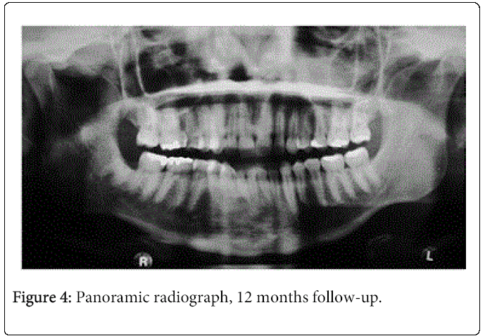
A 24-year-old female, came to our Oral Surgery Service in Dental Center of Studies and Research (COESP), João Pessoa, Paraíba, Brazil, in May 2012 for the extraction of lower third molars. An intentional partial odontectomy was performed on the tooth 48 in July 2012. The patient was satisfied without complaints about the loss of sensation and / or infection during 12 months follow up (Figures1, 2, 3(a-d) and 4).
Case 2
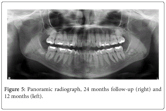
A 26-years-old female, came to our Oral Surgery Service in Dental Centre of Studies and Research (COESP), João Pessoa, Paraíba, Brazil, in May 2011 for the extraction of both lower third molars. The intentional partial odontectomy was performed in the tooth 48 in June 2011 and the tooth 38 in July 2012 with postoperative follow-up of 24 months and 12 months, respectively. The patient had not presented evidence of the loss of sensation and / or infection (Figures 5 and 6a-c).
Discussion
Several studies have demonstrated the success of the procedure of intentional partial odontectomy and these is emphatic in stating that it is a predictable and acceptable technique [1-4,6,11,12,14,16-18,19,21].
In fact, the intentional partial odontectomy presents itself as an alternative technique in extractions third molars which have a close relationship with the inferior alveolar nerve canal, as reported in present cases. Regarding the study of submerged roots with and without endodontic treatment, there is still no consensus in the literature because studies report good results in roots with endodontic treatment [5,10,16], while others disagree [8,9]. Reames et al. emphasizes that success in the roots with endodontic treatment has no predictability. The present cases did not have its roots endodontically treated.
Studies have verified the usefulness of computed tomography in preventing damage to the inferior alveolar nerve treated with both intentional partial odontectomy as due extraction of mandibular third molar with intimate relationship with the mandibular canal. Drage, Renton [12], Umar et al. [22] and Patel et al. [23] emphasized that the planned procedures such as CT scan, performed by trained surgeon and notion about the mechanical effect of surgical manipulation are important factors to decrease the permanent loss of sensitivity. We emphasize the CT was more accurate in diagnosing and predicting the exposure of the inferior alveolar neurovascular bundle for extraction of impacted mandibular teeth.
When it comes to the evaluation of the roots of mandibular third molars with proximity to the mandibular canal and if its removal, Howe, Poyton [11] and Rood, Shehab [24] found an incidence of sensory deficit in 35.64% and 30% of cases, respectively. When evaluated on panoramic radiographs, O'Riordan [1] found that when there was a radiolucent band across the root (defined as dark band on the root canal continuous white line), in 76% of cases were found injuries to the inferior alveolar nerve. When evaluated third molars treated by intentionally partial odontectomy, some studies have pointed found no damage to the inferior alveolar nerve [13,14,16,17,23]. while others reported the presence of transient paresthesias which resolved spontaneously on average one to three months postoperatively [15,22]. The present study was not observed any cases of temporary or permanent sensory deficit.
Studies revealed the rate of infection in roots remaining after intentional partial odontectomy was low [1,15,18,20,23] but only one study reported not having been any sign or symptom of postoperative infection [16]. When comparing the rate of postoperative infection in patients treated by intentional partial odontectomy and conventional extraction, indicate that the incidence of alveolitis was similar in both groups [13,15,18] In clinical cases presented no infection was observed until now.
Patel et al. [7] developed a study evaluating histologically 26 consecutive symptomaticroots in 21 patients. Found that all roots had vital tissue in the pulp chamber and there was no evidence of periradicular inflammation. Persistent postoperative symptoms related predominantly to inflammation of the soft tissue, which was caused by partially erupted roots or failure of the socket to heal.
In the analysis of the literature, no case had pain from the third month postoperatively [15,20]. The intentional partial odontectomy has a conservative nature, resulting in less disturbance to the tissue, thus postoperative pain can be reduced [23].
Based on the indicative images of periapical lesions and pathologies developed in the remaining roots, the studies do not indicate any associated injury exists after having been treated with the technique of intentional partial odontectomy [19,20].
Leizerovitz [2] reported a case report using the modified and grafted designed to minimize the drawbacks of standard. Modifications were: stabilizing the root stump to prevent intraoperative movement, creation of a large intrabony space for bone graft material, and grafting for periodontal healing while minimizing the possibility of postoperative root migration. In the same year, Leizerovitz,Leizerovitz3 published a case series using the modified and graftedSixteen patients, with total of 20 teeth, were followed for 6 to 49 months. Amendments to standard were intra-surgical stabilization of the root and the creation of a periodontal "scaffold." Modified and grafted showed excellent alveolar bone height and periodontal improvement, with remarkable bone regeneration. No residual root migration was evident on follow up, nor was there inadvertent intraoperative root removal. Suggest that the Modified and grafted may be considered a good alternative to a standard, especially in cases of high risk for, or existent periodontal defects on the distal of second molars or if no residual root migration is desired.
Ghaeminia [4] developed a study with randomised controlled trials (RCTs) and non-randomised controlled trials (CCTs) that comparedwith total removal for third molar extractions with high risk of nerve injury. Four studies (two RCTs and two CCTs) involving 699 patients and 940 third molars were included.was changed to total removal during surgery due to root loosening or mobilisation in 2.3% to 38.3% of cases. In 0% to 4.9% of cases reoperation was required in thegroup due to persistent pain, root exposure or persistent apical infections. Root migration was only reported in three studies and ranged from 13.2% to 85.9%. Suggest thatcan protect inferior alveolar nerves in the extraction of third molars with high risk of nerve injury as compared with total removal, and that the risk ratios of postoperative infections were similar between the two surgical modalities.
Conclusion
Given the present cases it can be concluded that:
•None of the reported cases was observed sensorial deficit and postoperative infection.
•Intentional partial odontectomy proved to be a predictable and effective technique which could be performed in an outpatient setting.
References
- O'Riordan BC (2004) Coronectomy (intentional partial odontectomy of lower third molars). Oral Surg Oral Med Oral Pathol Oral Radiol Endod 98: 274-280.
- Leizerovitz M,Leizerovitz O (2013) Modified and graftedcoronectomy: a new technique and a case report with two-year followup. Case Rep Dent 2013: 914173.
- Leizerovitz M,Leizerovitz O (2013) Reduced complications by modified and graftedcoronectomyvs. standardcoronectomy - a case series. Alpha Omegan 106: 81-89.
- Ghaeminia H (2013) Coronectomymay be a way of managing impacted third molars. Evid Based Dent 14: 57-58.
- Kim YB,Joo WH,Min KS (2014) Coronectomyof a lower third molar in combination with vital pulp therapy. Eur J Dent8: 416-418.
- Biocanin V,Todorovic L (2014) Coronectomyof two neighbouring ankylosed mandibular teeth - a case report. Vojnosanit Pregl71: 777-779.
- Patel V,Sproat C,Kwok J et al. (2014) Histological evaluation of mandibular third molar roots retrieved aftercoronectomy. Br J Oral Maxillofac Surg 52: 415-419.
- Whitaker DD, Shankle RJ (1974) A study of the histologic reaction of submerged root segments. Oral Surg Oral Med Oral Pathol 37: 919-935.
- Johnson DL, Kelly JF, Flinton RJ, Cornell MT (1974) Histologic evaluation of vital root retention. J Oral Surg 32: 829-833.
- Reames RL, Nickel JS, Patterson SS, Boone M, El-Kafrawy AH (1975) Clinical, radiographic, and histological study of endodontically treated retained roots to preserve alveolar bone. J Endod 1: 367-373.
- Howe GL, Poyton HG (1960) Prevention of damage to the inferior dental nerve during the extraction of mandibular third molars. Br Dent J 109: 355-363.
- Drage NA, Renton T (2002) Inferior alveolar nerve injury related to mandibular third molar surgery: an unusual case presentation. Oral Surg Oral Med Oral Pathol Oral Radiol Endod 93: 358-361.
- Renton T, Hankins M, Sproate C, McGurk M (2005) A randomised controlled clinical trial to compare the incidence of injury to the inferior alveolar nerve as a result of coronectomy and removal of mandibular third molars. Br J Oral Maxillofac Surg 43: 7-12.
- Pogrel MA (2007) Partial odontectomy. Oral Maxillofac Surg Clin North Am 19: 85-91.
- Leung YY, Cheung LK (2009) Safety of coronectomy versus excision of wisdom teeth: a randomized controlled trial. Oral Surg Oral Med Oral Pathol Oral Radiol Endod. 108: 821-827.
- Sencimen M, Ortakoglu K, Aydin C, Aydintug YS, Ozyigit A (2010) Is endodontic treatment necessary during coronectomy procedure? J Oral Maxillofac Surg. 68: 2385-2390.
- Leung YY, Cheung LK (2011) Risk factors of neurosensory deficits in lower third molar surgery: an literature review of prospective studies. Int J Oral Maxillofac Surg 40: 1-10.
- Cilasun U, Yildirim T, Guzeldemir E, Pektas ZO (2011) Coronectomy in patients with high risk of inferior alveolar nerve injury diagnosed by computed tomography. J Oral Maxillofac Surg. 69: 1557-1561.
- Goto S, Kurita K, Kuroiwa Y Hatano Y, Kohara K et al. (2012) Clinical and dental computed tomographic evaluation 1 year after coronectomy. J Oral Maxillofac Surg. 70: 1023-1029.
- Leung YY, Cheung LK (2012) Coronectomy of the lower third molar is safe within the first 3 years. J Oral Maxillofac Surg. 70: 1515-1522.
- Gleeson CF, Patel V, Kwok J, Sproat C (2012) Coronectomy practice. Paper 1. Technique and trouble-shooting. Br J Oral Maxillofac Surg 50: 739-744.
- Umar G, Obisesan O, Bryant C, Rood JP (2013) Elimination of permanent injuries to the inferior alveolar nerve following surgical intervention of the "high risk" third molar. Br J Oral Maxillofac Surg 51: 353-357.
- Patel V, Gleeson CF, Kwok J, Sproat C (2013) Coronectomy practice. Paper 2: complications and long term management. Br J Oral Maxillofac Surg 51: 347-352.
- Rood JP, Shehab BA (1990) The radiological prediction of inferior alveolar nerve injury during third molar surgery. Br J Oral Maxillofac Surg 28: 20-25.
Relevant Topics
- Advanced Bleeding Gums
- Advanced Receeding Gums
- Bleeding Gums
- Children’s Oral Health
- Coronal Fracture
- Dental Anestheia and Sedation
- Dental Plaque
- Dental Radiology
- Dentistry and Diabetes
- Fluoride Treatments
- Gum Cancer
- Gum Infection
- Occlusal Splint
- Oral and Maxillofacial Pathology
- Oral Hygiene
- Oral Hygiene Blogs
- Oral Hygiene Case Reports
- Oral Hygiene Practice
- Oral Leukoplakia
- Oral Microbiome
- Oral Rehydration
- Oral Surgery Special Issue
- Orthodontistry
- Periodontal Disease Management
- Periodontistry
- Root Canal Treatment
- Tele-Dentistry
Recommended Journals
Article Tools
Article Usage
- Total views: 20974
- [From(publication date):
March-2015 - Aug 20, 2025] - Breakdown by view type
- HTML page views : 15870
- PDF downloads : 5104

