Case Report Open Access
Dentigerous cyst Associated with Compound Odontome
Sachin A Gunda1, Ankita R Hingmire1, Anil T Patil1*, Anand L Shigli1 and Dilip Magdum2
1Pedodontics and Preventive Dentistry, Department of Pedodontics and Preventive Dentistry, Bharati Vidyapeeth Deemed University Dental College and Hospital, Sangli, Maharashtra, India
22Department of Oral Pathology, Bharati Vidyapeeth Deemed University Dental College and Hospital, Sangli, Maharashtra, India
- *Corresponding Author:
- Patil AT
Department of Pedodontics and Preventive Dentistry
Bharati Vidyapeeth University Dental College and Hospital
Sangli 416414, India
Tel: +91 9850983500
E-mail: dranilp0888@gmail.com
Received Date: September 19, 2016; Accepted Date: October 20, 2016; Published Date: October 28, 2016
Citation: Gunda SA, Hingmire AR, Patil AT, Shigli AL, Magdum D (2016) Dentigerous cyst Associated with Compound Odontome. Pediatr Dent Care 1: 121.
Copyright: © 2016 Gunda SA, et al. This is an open-access article distributed under the terms of the Creative Commons Attribution License, which permits unrestricted use, distribution, and reproduction in any medium, provided the original author and source are credited.
Visit for more related articles at Neonatal and Pediatric Medicine
Abstract
The most common of the odontogenic tumours of the jaws are odontomas. Occasionally, a dentigerous cyst might develop from epithelial lining of fibrous capsule of a odontoma. We present a case of a 10-year-old boy suffering from apparent ectopic partial eruption of mandibular right first premolar associated with an impacted compound odontome related with a dentigerous cyst.
Keywords
Compound Odontome; Dentigerous cyst; Oral
Introduction
The most common of the odontogenic tumors of the jaws are odontomas. They are mixed tumors, comprised of both mesenchymal and epithelial cells that present a complete dental tissue differentiation (enamel, dentin, cementum and pulp) [1]. Odontomas are defined as nonaggressive hamartomatous developmental malformations or lesions of odontogenic origin which consists of enamel, dentin, cementum and pulpal tissue by the World Health Organization (WHO). Odontomas are of two types namely complex odontomas and compound odontomas. Complex odontomas are malformation where dental tissues are present, but arranged in a more or less disorderly pattern; and compound odontomas are malformation having all dental tissues in a pattern that is more orderly arranged. Etiology is still unknown of these hamartomas odontomas [2]. Large odontomas may cause delayed eruption and cystic development [1]. Dentigerous cysts are always associated with a developing or unerupted tooth bud [3]. Less frequently, they can be found in relation with supernumerary teeth, odontomas or unerupted deciduous teeth. Dentigerous cyst is rarely seen associated with an erupted tooth [4]. Herein we have documented a case of dentigerous cyst associated with compound odontome in the mandibular left first premolar region.
Case Report
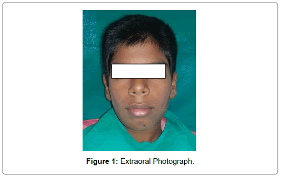
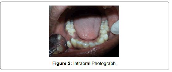
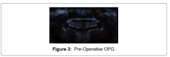
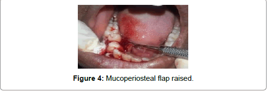
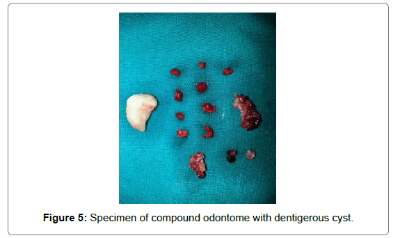
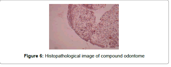
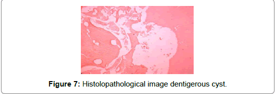
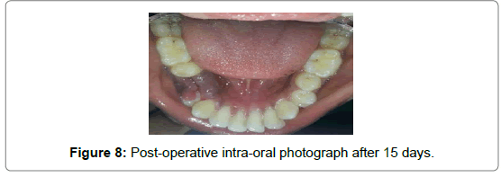
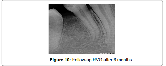
A 10-year-old boy reported to the pedodontics department of the Bharati Vidyapeeth Dental College & Hospital, Sangli with chief complaint of malaligned right lower back tooth. Patient’s mother gave history of pain and swelling associated with the same tooth one month back. The pain was mild in nature associated with intra-oral swelling. The family history and medical histories were non-contributory. No history of trauma to the face or mouth was recalled. Extra-oral examination was non-significant (Figure 1). Intra-oral examination revealed mixed dentition period, rotated right mandibular second premolar and buccally erupting right mandibular first premolar (Figure 2). No inflammatory signs were noted in the gingiva and alveolar bone but a bulge appeared on the buccal surface in the right lower premolar region (Figure 2). A panoramic radiographic examination showed an unerupted supernumerary tooth. A well demarcated unilocular radiolucent follicle occupied by a radio-opaque mass was observed (Figure 3). The overlying alveolar bone was normal. The provisional diagnosis was a para-premolar, which lead to ectopic eruption of the first premolar. A full thickness mucoperiosteal flap was reflected buccally between the right mandibular first and second premolar. Surgical removal of the odontome and the cyst lining was accomplished under local anesthesia. A thin layer of bone over the buccal surface was removed and the tooth like structure was exposed (Figure 4); which was removed along with the cystic lining (Figure 5). The extraction cavity was curreted and was devoid of granulation tissue. The flap was replaced and secured with sutures. Histopathologically, the single rooted tooth like structure was exhibiting normal dentinal tubules, predentin and pulpal tissue was observed. Hence histopathological evaluation confirmed it to be a compound type of odontome (Figure 6). The biopsy specimen was sent to the pathology lab for investigation which was confirmed as ‘dentigerous cyst’ (Figure 7). The post-operative period was uneventful (Figures 8-10). The patient is being monitored at regular intervals. This case shows a compound odontoma evidenced on radiograph by multiple denticles encircled and a radiolucent area in the right first premolar region of mandible associated a dentigerous cyst.
Discussion
Odontomas are hamartomas made up of enamel, dentin, cementum and occasionally pulp. They are non-aggressive in behavior and are slow growing benign tumors. They are classified as compound if there is superficial anatomic similarity to even rudimentary teeth and complex, when the calcified tissue present as an irregular mass composed mainly of mature tubular dentin. Compound odontomas are more common than complex and are present in the ratio of 2:1. Complex odontomas are seen in the posterior region of jaw and compound odontomas are seen more in the anterior maxilla. Clinically odontomas can be intraosseous and extra-osseous. Intra–osseous odontomas occur inside the bone and may erupt. Extra-osseous odontomas (erupted odontomas) found in soft tissue covering the tooth bearing portion of the jaws. The odontomas are found commonly in the first two decades of life, with no gender preferences. Odontomas are usually asymptomatic but clinical signs can be seen like unerupted permanent teeth, pain, over-retained deciduous teeth, tooth displacement and expansion of cortical bone. Anesthesia in the lower lip and swelling in the affected region are the other symptoms.
Odontomas have density that is more than that of bone and equal to or greater than that of tooth and is seen as a well-defined radioopacity in bone, which contains foci of variable density. A radiolucent halo, peculiarly surrounded by thin sclerotic line, surrounds the radio-opacity. The radiolucent zone is the connective tissue capsule of a normal tooth follicle [5]. The compound odontoma was defined as an injury that stems from an exaggerated proliferation of the dental lamina, showing all dental tissues in an orderly manner and are usually presented in the form of denticles. Radiographically compound odontoma are a group of structures similar to the teeth can have different sizes and shapes, encircled by a radiolucent area. In the complex odontoma, dental tissues are found like an amorphous calcified mass and radiographically observed as uniform radiopacity with irregular edges surrounded by radiolucent space [6,7]. Odontomas are inherited through a mutant gene or interference, possibly post natal, with genetic control of tooth development was suggested by Hitchin. As a consequence of their odontogenic nature, including epithelial and mesenchymal tissue odontomas can form cystic transformation into dentigerous cyst. It results from the cystic degeneration of enamel organ after partial or complete crown development, cystic transformation of the follicle associated with the unerupted tooth may also happen when its eruption is impeded by the odontoma [5]. The follicular cyst, which is also called as a dentigerous cyst are true developmental cysts predominantly affecting mandibular third molars of young patients, followed by maxillary canine, mandibular premolar and maxillary third molar.
Dentigerous cyst associated with an unerupted compound odontome is an extremely rare entity which has been reported in our case. Dentigerous cyst is usually asymptomatic and can be associated with mobility or displacement of teeth, radiographically characterized by a radiolucent lesion with a well-defined sclerotic margin associated with a crown of an unerupted tooth. Radiographically, three variants of dentigerous cysts namely central, lateral and circumferential have been described. A large dentigerous cyst may appear multilocular radiographically owing to the persistence of bone trabeculae within the radiolucency [4]. Histopathologically it comprises of a fibrous conjunctive tissue capsule and an epithelial lining of flattened cells with either presence or absence of keratinization. Complications related to dentigerous cysts are pathological bone fracture, loss of permanent teeth, bone deformities and development of ameloblastoma or malignancies such as squamous cell carcinoma and intra-osseous mucoepidermoid carcinoma.
The treatment of odontoma is surgical excision, which has great success with low recurrence rare. The treatment for dentigerous cysts is surgical excision with removal of the involved tooth, in few cases marsupialization of the lesion is needed to reduce bone defect before surgical intervention [6]. The surgical exposure showed the cystic lesion attached to the neck of the unerupted tooth. Hence it was treated by surgical excision of the lesions and the associated tooth. The size of the lesion did not require marsupialization before removal and a satisfactory amount of surrounding bone was preserved.
Conclusion
The compound odontoma is benign malformation usually asymptomatic, with slow evolution and may be associated with other disorders such as dentigerous cyst as outlined in this case. The treatment of choice is surgical excision of the lesions along with the toothassociated to the cyst. Surgical excision of odontome with cyst in children should be performed with careful, imperative, immediate planning; preventing injury to vital structures and developing occlusion.
References
- Sales MG (2009) Cavalcanti. Complex odontoma associated with dentigerous cyst in maxillary sinus: case report and computed tomography features. Dentomaxillofacial Radiology 38: 48-52.
- Amailuk P, Grubor D (2008) Erupted compound odontoma: case report of a 15-year-old Sudanese boy with a history of traditional dental mutilation.British Dental Journal,204: 11-14.
- Gulses A, Karacayli U, Koymen R (2009) Dentigerous Cyst Associated With Inverted and Fused Supernumerary Teeth in a Child: A Case Report. OHDMBSC 8: 1-4.
- Shergill AK, Singh P, SolomonMC, Singh PG (2014)Dentigerous Cyst Associated with an EruptedTooth – An Unusual Presentation. International Journal of Scientific Study 2: 1-3.
- Shah A, Singh M, Chowdhury S (2010) Complex Odontoma associated with Dentigerous cyst.Int Journal Of Clinical Dental Science 1: 1-4.
- Melo RB, Damasceno, Celio YES,Couto da Cunha Junior A, PontesIV(2015) Compound odontoma associated with dentigerouscyst in the anterior mandible -Case Report. RSBO 12: 98-102.
- Tomizawa M, Otsuka Y, Noda T (2005) Clinical observations of odontomas in Japanese children: 39 cases including one recurrent case.Int J Paediatr Dent 15: 37-43.
Relevant Topics
- About the Journal
- Birth Complications
- Breastfeeding
- Bronchopulmonary Dysplasia
- Feeding Disorders
- Gestational diabetes
- Neonatal Anemia
- Neonatal Breastfeeding
- Neonatal Care
- Neonatal Disease
- Neonatal Drugs
- Neonatal Health
- Neonatal Infections
- Neonatal Intensive Care
- Neonatal Seizure
- Neonatal Sepsis
- Neonatal Stroke
- Newborn Jaundice
- Newborns Screening
- Premature Infants
- Sepsis in Neonatal
- Vaccines and Immunity for Newborns
Recommended Journals
Article Tools
Article Usage
- Total views: 13477
- [From(publication date):
specialissue-2016 - Aug 17, 2025] - Breakdown by view type
- HTML page views : 12425
- PDF downloads : 1052

