Research Article Open Access
Discerning Viable from Nonviable Yersinia Pestis pgm- and Bacillus Anthracis Sterne using Propidium Monoazide in the Presence of White Powders
Becky M. Hess*, Brooke L. Deatherage Kaiser, Michael A. Sydor, David S. Wunschel, Cynthia J. Bruckner-Lea and Janine R. Hutchison
Chemical and Biological Signature Sciences, Pacific Northwest National Laboratory, Richland, Washington, United States of America
- *Corresponding Author:
- Becky M. Hess
Pacific Northwest National Laboratory
902 Battelle Boulevard, P.O. Box 999
MSIN: P7-50, Richland, WA 99352, USA
Tel: 509-372-6792
Fax: 509-375-2227
E-mail: Becky.Hess@pnnl.gov
Received Date: August 05, 2015; Accepted Date: November 20, 2015; Published Date: November 26, 2015
Citation: Hess BM, Kaiser BLD, Sydor MA, Wunschel DS, Lea CJB, et al. (2015) Discerning Viable from Nonviable Yersinia Pestis pgm- and Bacillus Anthracis Sterne using Propidium Monoazide in the Presence of White Powders. J Bioterror Biodef 6:138 doi: 10.4172/2157-2526.1000138
Copyright: © 2015 Hess BM, et al. This is an open-access article distributed under the terms of the Creative Commons Attribution License, which permits unrestricted use, distribution, and reproduction in any medium, provided the original author and source are credited.
Visit for more related articles at Journal of Bioterrorism & Biodefense
Abstract
Purpose of the study To develop and optimize an assay to determine viability status of Bacillus anthracis Sterne and Yersinia pestis pgm- strains in the presence of white powders by coupling propidium monoazide (PMA) treatment with real-time PCR (qPCR) analysis. Approach and results After gaining entry to intracellular space, PMA can be exported by metabolically active cells. The PMA remaining in nonviable cells binds DNA, thereby increasing qPCR assay cycle threshold (CT) values compared to untreated samples. Dye concentration, cell number and fitness, incubation time, inactivation methods, and assay buffer were optimized for a Gram-positive pathogen, B. anthracis Sterne, and a gram negative pathogen, Y. pestis pgm-. Differences in CT values in nonviable cells compared to untreated samples were consistently > 9 for both B. anthracis Sterne vegetative cells and Y. pestis pgm- in the presence and absence of three different white powders. Our method eliminates the need for a DNA extraction step prior to detection by qPCR. Significance of the Study The method developed for simultaneous detection and viability assessment for B. anthracis and Y. pestis can be employed in forming decisions about the severity of a bio threat event or the safety of food.
Keywords
Bacillus anthracis; Yersinia pesti; Propidium monoazide; QPCR; White powders; Rapid viability detection
Introduction
Bacillus anthracis is a spore-forming, Gram-positive bacterium and is the causative agent of the zoonotic disease anthrax. Infection occurs when spores gain access to the host through respiratory, cutaneous, or gastrointestinal routes [1]. B. anthracis is listed by the CDC as a category A bio threat agent because of the stability of the spore form, infectivity, and ease of dissemination. In 2001, the intentional dissemination of B. anthracis spores through the mail prompted public health officials and the scientific community to improve methods for rapid detection of bio threat agents in general [2]. Of additional concern, Y. pestis is also a category a bio threat agent and is the causative agent of plague [3,4]. Given the bio threat potential of both Y. pestis and B. anthracis, we sought to develop a rapid viability determination method for these two organisms [4-7].
The need for rapid methods to detect DNA signatures from suspected bio threat events has largely been met with real-time PCR (qPCR) methods, which can be used to quickly identify bacterial species using specifically designed probes. However, qPCR assays alone do not assess bacterial viability [8-12], which is critical information to ensure an appropriate and timely response to bio threat events. While many assays have been developed to assess bacterial viability for certain types of bacterial cells, bacterial spores remain challenging due to their dormant nature but infectious potential.
Published work has studied the effectiveness of determining viable from nonviable samples by pretreating with intercalating dyes propidium monoazide (PMA) or ethidium monoazide (EMA) prior to conducting qPCR assays. PMA enters cells and cannot be actively exported from nonviable cells or cells with damaged membranes and forms covalent bonds with DNA after photo activation [13-15]. Previous work by Nocker et al. showed that EMA is capable of penetrating live as well as dead cells; therefore, EMA was not used in our study [14]. The binding action (PMA-DNA covalent bond) renders the bound DNA inaccessible to the polymerase and therefore undetectable in the qPCR reaction, thereby increasing the CT value compared to equivalent untreated samples [12,16]. Previous studies using intercalating dyes coupled with qPCR (i.e., PMA-qPCR ) detection have focused on bacterial species commonly associated with food-borne illnesses, such as Escherichia coli, Listeria monocytogenes, Salmonella enterica serovar Typhimurium, and Campylobacter jejuni, in which artificially spiked foods were subjected to PMA treatment followed by qPCR to determine the presence and viability of the bacteria. Ultimately, the conclusions of these studies indicate that PMA coupled qPCR is a promising tool to identify viability, presence, and species, but that assay optimization is required for each system [12,16-21]. The purpose of the current study is to assess the suitability of this method for bio threat agents Y. pestis and B. anthracis, and to define assay conditions and critically test the method under laboratory conditions. We found that the assay is well suited to discerning viable from nonviable cells of both of the tested bacteria in a laboratory setting and in the presence of three different white powders. A DNA extraction is not required, thereby streamlining the process and reducing labor requirements compared to other published methods. Therefore, the PMA-qPCR assay described here is a promising method for rapid detection and viability assessment of B. anthracis Sterne and Y. pestis pgm- vegetative cells in a controlled, laboratory setting and in the presence of the white powders included in this study.
Materials and Methods
Bacterial strains and culture conditions
Yersinia pestis KIM D27 (pgm-) and vegetative Bacillus anthracis Sterne were grown by selecting a fresh colony from a Tryptic Soy Agar (TSA; BD Biosciences, Franklin Lakes, New Jersey) streak plate derived from glycerol stocks. The colony was used to inoculate fresh tryptic soy broth without dextrose (TSB; BD) in a glass flask (50 mL culture volume for Y. pestis pgm- and 10 mL culture volume for B. anthracis Sterne). Overnight cultures were grown at 37°C (B. anthracis Sterne) or 30°C (Y. pestis pgm-) at 200 rpm for 16 to 18 hours. Log phase B. anthracis Sterne cultures were prepared by adding 1 mL of overnight culture to 9 mL of fresh TSB in a 50 mL glass flask and incubating at 37°C and 200 rpm for three to four hours. Log phase Y. pestis pgmcultures were prepared by adding 15 mL of overnight cultures to 15 mL of fresh TSB and incubating at 30°C and 200 rpm for four to five hours.
Bacillus anthracis Sterne spore stocks were generated using nutrient broth supplemented with CCY salts as described by Buhr [22]. Briefly, B. anthracis Sterne was inoculated into TSB at 37°C and agitated for 16 to 18 hours at 200 rpm. The sporulation media was then inoculated (1:100) using the overnight culture and incubated at 37°C, 200 rpm for 72 hours. The suspension was then centrifuged for 10 minutes at 10,000 × g and the resulting supernatant was discarded. The biomass was suspended in 30 mL of sterile, milli Q water and stored at 4°C for seven days to promote vegetative cell lysis. The sample was then washed three times with 30 mL of sterile, milliQ water by centrifugation for 10 minutes at 10,000 × g and resuspended in a final volume of 10 mL. The spores were visualized using phase contrast microscopy and > 95% of observed spores was phase-bright. The spore stocks were maintained at 4°C for the duration of the study. All bacterial stocks were enumerated prior to conducting experiments by plating serial dilutions in PBS (Life Technologies, Carlsbad, CA) containing 0.02% Tween 80 (Sigma Aldrich, St. Louis, MO) on TSA plates at the appropriate temperature.
Generating a template for qPCR reactions by heat lysis or DNA extraction
DNA extractions were performed using the Qiagen DNeasy Blood and Tissue Kit according to the manufacturer’s instructions, without any deviations. As described in the qPCR section below, direct heat lysis was conducted by including a single cycle at the beginning of the qPCR method at 95°C for ten minutes.
Sample inactivation
Y. pestis pgm- was inactivated using 50% ethanol. Aliquots of log phase or overnight cultures were centrifuged at 16,000 x g for 5 minutes and resuspended in the equivalent volume of 50% ethanol (prepared fresh just before use by diluting 200 proof ethanol at a 1:1 ratio with nuclease free water) and transferred to a new tube. Samples were incubated at room temperature for 30 minutes. Half of the sample volume was plated on TSA plates and incubated for 48 hours at 30°C to verify lack of cell growth and inactivation. The B. anthracis Sterne spores and vegetative cells were inactivated using 3% hydrogen peroxide (Sigma Aldrich) (prepared fresh just before use by diluting a 35% stock of hydrogen peroxide with nuclease free water). Aliquots of log phase culture, overnight cultures, and up to 109 spores were centrifuged at 16,000 × g for 5 minutes and resuspended in the equivalent volume of 3% hydrogen peroxide. Samples were incubated at room temperature for one hour. For all experiments, half of the sample was plated on TSA plates and incubated for 48 hours at 37°C to verify inactivation; all samples were negative for bacterial growth. Following the indicated exposure time to the inactivation agent, the remaining sample volume was centrifuged at 16,000 × g for five minutes and the supernatant was then discarded. The resulting sample was resuspended in the equivalent volume of Tris-EDTA pH 8 Buffer (TE, Ambion, Carlsbad, CA) and transferred to a new tube.
Addition of white powders to samples
The white powders were purchased from a local vendor. We selected Peak Dry whole milk powder (referred to in the text as milk powder), Rumford aluminum free baking powder (referred to in the text as baking powder), and Coffee-mate “The Original” coffee creamer (referred to in the text as coffee creamer). Suspensions of each powder were prepared in TE at a stock concentration of 5 mg/mL. To ensure the white powder did not contribute to viable biomass measurements, the powder suspensions were sterilized by autoclaving for 20 minutes on a liquid cycle at 121°C. Sterilization was confirmed by plating 100 μL aliquots on TSA plates and confirming lack of growth after 48 hours at 37°C. Samples containing white powder were prepared as described above; however, the final resuspension of the sample was conducted using fresh TE spiked with the white powder suspension to a final concentration of 0.1 mg/mL white powder in TE.
Preparing and treating samples with PMA
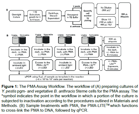
All dilutions were performed using TE buffer. The indicated concentration of PMA was added to 500 μL aliquots of viable or nonviable cells. An assay workflow is depicted in (Figure 1). The 20 mM PMA stock (Biotium, Hayward, CA) was stored at -20°C and diluted to the required concentrations using nuclease free water prior to use. Sample volumes were 500 μL and we prepared concentrations of PMA such that the addition of 1.25 μL to the sample would result in the desired PMA concentration. Samples were incubated in the dark and covered with aluminum foil for five minutes at room temperature. Following the incubation, the samples were photo activated using the PMA-LiteTM LED Photolysis Device (Biotium) as the LED sources to photo activate the PMA for five minutes (Figure 1B).
Figure 1: The PMA Assay Workflow. The workflow of (A) preparing cultures of Y. pestis pgm- and vegetative B. anthracis Sterne cells for the PMA assay. The *symbol indicates the point in the workflow in which a portion of the culture is subjected to inactivation according to the procedures outlined in Materials and Methods. (B) Sample treatments with PMA, the PMA-LITETM which functions to cross-link the PMA to DNA, followed by qPCR.
Quantitative PCR (qPCR) experiments
Primer sets are provided in (Table 1). Y. pestis primers and probes targeted the chromosomal gene yihN as described by Stewart et al. [3]. B. anthracis primer and probe targeted the CAAX gene as described by Wielinga et al. [23]. Following PMA treatment, 5 μL of sample was used as the template in the qPCR reaction. Each qPCR reaction was performed in a 20 μL volume with the following reaction components: 10 μL of 2x TaqMan® Fast Universal Master Mix (Life Technologies), 1 μL of 20x Primer/Probe (PrimeTime® qPCR Assays; IDT, Coralville, IA), 4 μL of nuclease free water, and 5 μL of template. TE was used as the template for the no template control reactions. Each sample was run in triplicate, and the experiments were conducted a total of three times independently (n=9). PCR was conducted using the Applied Biosystems 7500 Real-Time PCR system with the default fast platform settings (95°C for ten minutes for heat lysis, 95°C for 10 s, followed by 40 cycles of denaturation at 95°C for 3 s and annealing/extension at 60°C for 30 s). CT values greater than 35 were considered as undetected by the qPCR assay, abbreviated as ND in the presented data. A workflow of the full assay is depicted in (Figure 1).
| Target | Primer Name | Sequence(5’ – 3’) |
|---|---|---|
| B. anthracischromosome | CAA × _F | TCC GTT TAC CAA TTC ACT ATG AAT CAA T |
| CAA × _R | ATG CGT TGT TAA GTA TTG GTA TAA TCA TC | |
| CAA × _ Probe |
FAM/CC CAC TTG G/Zen/A TTA TAT CCT GAG TAT CGT GA/3IABkFQ/ | |
| Y. pestischromosome | Yp_F | CGC TTT ACC TTC ACC AAA CTG AAC |
| Yp_R | GGT TGC TGG GAA CCA AAG AAG A | |
| Yp_ Probe |
FAM/TA AGT ACA T/Zen/C AAT CAC ACC GCG ACC CGC TT/3IABkFQ/ |
Table 1: Primers and probes used in this study.
The difference in CT values was used to compare nonviable samples with viable samples to the appropriate non-treatment control using the following formulas:
1) ΔCT = ΔCTnonviable – ΔCTviable,
2) ΔCTnonviable = CT nonviable with PMA– CT nonviable without PMA, and
3) ΔCTviable = CT viable with PMA– CT viable without PMA.
Quantitation
As a reference method for quantitation of the bacteria, after culture preparation, but prior to PMA or LED treatment, aliquots of bacterial samples were serially diluted and plated on TSA plates to determine total cell numbers in all qPCR reactions. These values could then be correlated back to raw CT values obtained in the experiments to ensure consistent biomass in all treatment samples and qPCR reactions. The number of cells in each qPCR reaction is provided in the figure legends for each relevant data set. In this way, the CT value can be used to deduce the amount of bacteria in unknown samples.
Statistical analysis
Statistical analysis was conducted using the student’s one-tail t-Test with the Analysis Tool Pak Excel add-in tool. Results were considered significant if the resulting p value was less than or equal to 0.05. The data set for each t-Test consisted of a minimum of three biological replicates for each sample type. That is, three independent experiments were conducted for each sample type. In each independent experiment, a total of three technical replicates were conducted (n=9).
Results
Evaluating PMA treatment with Y. pestis pgm-, B. anthracis sterne vegetative cells, and B. anthracis sterne spores
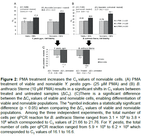
We first wanted to test if populations of cells could be simultaneously identified and determined as viable or not without performing a DNA extraction. To test this method, we subjected equivalent quantities of viable and nonviable cells or spores to PMA treatment and the PMALite TM according to the procedures outlined in Materials and Methods and diagrammed in (Figure 1). We empirically determined the optimal incubation time, assay buffer, inactivation method, LED exposure time, and growth phase for B. anthracis Sterne and Y. pestis pgm- (data not shown). As shown in (Table 2), there was a shift in the CT values between the viable and nonviable cell populations for both Y. pestis pgm- and B. anthracis Sterne vegetative cells. For clarity, a workflow of data analysis from raw CT values to ΔCT for the vegetative cells from this data set is shown in (Figure 2). For both cell types, qPCR detection was successful without a DNA extraction (untreated and treated). The optimized parameters for the assay are described in (Table 3).
| Organism | Viability | Untreated CT (SD) | LED CT (SD) | LED + PMA CT (SD) | ΔCT viable or ΔCT nonviable | ΔCT |
|---|---|---|---|---|---|---|
| B. anthracisSterne spores | Nonviable | 31.87 (0.30) | 32.17 (0.22) | ND | N/A | N/A |
| B. anthracisSterne spores | Viable | 33.67 (0.16) | 34.25 (0.31) | ND | N/A | |
| B. anthracisSterne Vegetative | Nonviable | 21.76 (0.07) | 21.80 (0.11) | 34.15 (0.40) | -12.4 | -7.2 |
| B. anthracisSterne Vegetative | Viable | 21.66(0.03) | 21.87 (0.18) | 26.90 (0.24) | -5.24 | |
| Y. pestispgm- | Non-viable | 16.1 (0.06) | 16.1 (0.17) | 27.5 (1.03) | -11.4 | -6.6 |
| Y. pestispgm- | Viable | 16.6 (1.47) | 16.6 (1.46) | 21.4 (1.19) | -4.8 |
Table 2: PMA treatment distinguishes viable from nonviable B. anthracis Sterne vegetative cells and Y. pestis pgm- cells but not B. anthracis spores. Raw CT values of of viable and nonviable samples. The PMA concentration is 10 μM for B. anthracis Sterne spores and vegetative cells and 25 μM for Y. pestis pgmsamples. Spore samples contained 15,000 spores. ND, absence of qPCR signal. N/A, not applicable. Measurements are the average value of three independent e × periments. All ΔCT values are calculated using equations (1), (2), or (3) as described in Materials and Methods.
| Variable | Y. pestis pgm- | B. anthracis Sterne |
|---|---|---|
| Cell state | Log phase | Log phase, vegetative cells |
| Cell number in qPCR reaction | 5 × 10³ to 107 | 5 × 10³ to 107 |
| Inactivation Method | 50% ethanol exposure for 30 minutes | 3% H2O2exposure for 60 minutes |
| PMA Concentration | 25μM | 10μM |
| PMA Pretreatment Time | 5 minutes | 5 minutes |
| Exposure to PMA-LiteTM | 5 minutes | 5 minutes |
| Assay Buffer | TE | TE |
Table 3: Summary of optimized conditions for each model system used in the study, compatible with 0.1 mg/mL concentration of white powders discussed in the te × t.
Figure 2: PMA treatment increases the CT values of nonviable cells. (A) PMA treatment of viable and nonviable Y. pestis pgm- (25 μM PMA) and (B) B. anthracis Sterne (10 μM PMA) results in a significant shifts in CT values between treated and untreated samples (ΔCT). (C)There is a significant difference between the ΔCT values of viable and nonviable cells, enabling differentiation of viable and nonviable populations. The *symbol indicates a statistically significant difference (p < 0.05) when comparing the ΔCT values of viable and nonviable populations. Among the three independent experiments, the total number of cells per qPCR reaction for B. anthracis Sterne ranged from 3.1 × 10⁶ to 3.8 × 10⁶ which corresponded to CT values of 21.66 to 21.76. For Y. pestis, the total number of cells per qPCR reaction ranged from 5.9 × 10⁵ to 6.2 × 10⁵ which corresponded to CT values of 16.1 to 16.6.
To ensure that the genomic DNA extraction step could be eliminated from the assay workflow, we compared the qPCR signal from viable and nonviable bacteria under our culture and experimental conditions using direct heat lysis and DNA extraction using a Qiagen DNeasy Blood and Tissue kit. The genomic DNA was extracted according to the manufacturer’s instructions without deviations. Both Y. pestis pgmand B. anthracis Sterne were grown under conditions described in (Figure 1A). The resulting cultures were centrifuged and split into three equivalent aliquots. Two of the aliquots were designated as viable and a third was subjected to inactivation methods described in the Materials and Methods. One viable sample was subjected to DNA extraction and resuspended in an equivalent volume as the untreated samples. All samples were then analyzed by qPCR. The CT value in viable Y. pestis pgmd- for the extracted DNA was 15.98 ± 0.09; compared to untreated samples which had CT values of 16.2 ± 0.02 for the viable sample and 16.41 ± 0.03 for the nonviable sample. Similar results were achieved with B. anthracis Sterne. Extracted DNA from viable B. anthracis Sterne had CT value of 18.82 ± 0.02; compared to 19.11 ± 0.01 for the untreated viable sample and 19.19 ± 0.04 for the untreated nonviable sample. Given these results, we determined that the difference between CT values was not significant in extracted DNA versus cells lysed by direct heat lysis [24] and that a DNA extraction would not be required for either organism used in our study. Lending further support to our decision to eliminate the DNA extraction step can be found in the work done by Nocker et al. which showed that excess PMA (i.e., PMA that did not interact with DNA molecules) is inactivated by reacting with water molecules in solution, prior to DNA extraction, and thus will not affect the DNA from viable cells after cell lysis [14,15].
B. anthracis Sterne spores are expected to be coated in chromosomal DNA because of the interaction with the chromosome of the mother cell which gives rise to the spore [25-27]. For this reason, we postulated that treating spores with PMA would result in the PMA binding to the DNA coating the outside of the spore rather than penetrating to the interior of the spore, and would result in the absence of a qPCR signal. Further, the robust nature of the spore structure would limit the amount of PMA able to enter the spore. Therefore, we expected that treating both viable and nonviable spores with PMA would yield the same result: an absence of a qPCR signal and an inability to differentiate viable from nonviable spores. To test this idea, spores were inactivated using 3% hydrogen peroxide as described in the Materials and Methods to generate nonviable spores. The same number of viable and nonviable spores in each independent experiment were treated with 10 μM PMA with or without five minute exposure to LED light prior to qPCR detection. As shown in (Table 2), we obtained the postulated results for viable and nonviable spores; there is no difference in the qPCR results from viable and nonviable samples. Therefore, we concluded that this assay is not compatible with spores under the tested conditions; spore samples would require germination to a vegetative cell state in order to be compatible with this assay.
Optimizing PMA concentrations for cells
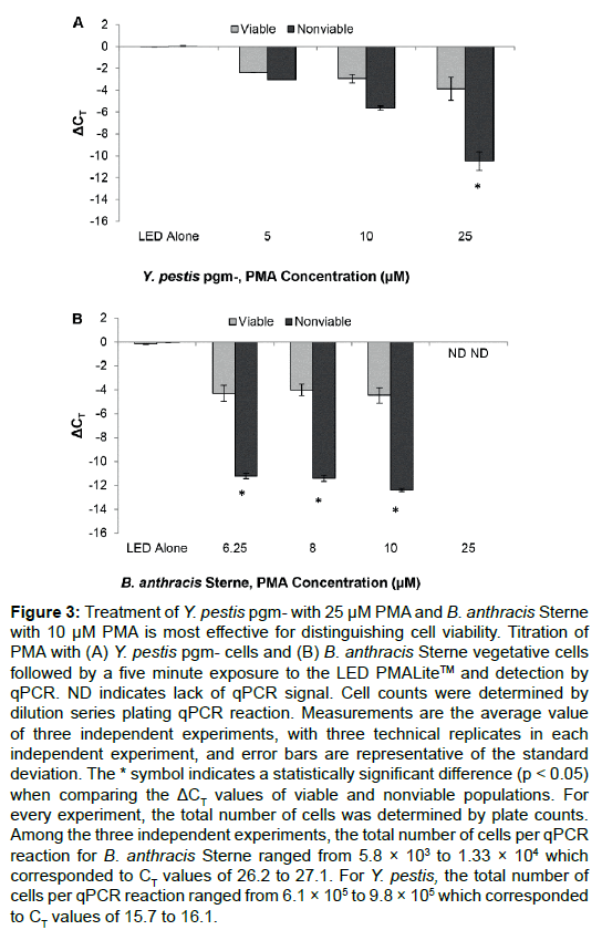
To find the most effective PMA concentration for Y. pestis, we tested concentrations of PMA ranging from 5 μM to 25 μM. The number of cells in each sample ranged from 5 × 10³ to 1 × 10⁶ CFU/ μL, with most samples containing 104 CFU/μL (i.e., approximately 5 × 10⁴ cells per qPCR reaction per 5 μL of sample used in the qPCR reaction). For samples treated with light alone, or PMA alone, there was not a significant difference in CT values between the viable and nonviable cells (Figure 3A). PMA treatment at 25 μM concentration resulted in the greatest change in magnitude of the PCR signal and was therefore selected as the optimal concentration for Y. pestis.
Figure 3: Treatment of Y. pestis pgm- with 25 μM PMA and B. anthracis Sterne with 10 μM PMA is most effective for distinguishing cell viability. Titration of PMA with (A) Y. pestis pgm- cells and (B) B. anthracis Sterne vegetative cells followed by a five minute exposure to the LED PMALiteTM and detection by qPCR. ND indicates lack of qPCR signal. Cell counts were determined by dilution series plating qPCR reaction. Measurements are the average value of three independent experiments, with three technical replicates in each independent experiment, and error bars are representative of the standard deviation. The * symbol indicates a statistically significant difference (p < 0.05) when comparing the ΔCT values of viable and nonviable populations. For every experiment, the total number of cells was determined by plate counts. Among the three independent experiments, the total number of cells per qPCR reaction for B. anthracis Sterne ranged from 5.8 × 10³ to 1.33 × 10⁴ which corresponded to CT values of 26.2 to 27.1. For Y. pestis, the total number of cells per qPCR reaction ranged from 6.1 × 10⁵ to 9.8 × 10⁵ which corresponded to CT values of 15.7 to 16.1.
With respect to B. anthracis Sterne cells, we observed consistent ΔCT values in viable and nonviable populations with PMA concentrations between 6.25 μM up to 10 μM (Figure 3B). Neither viable nor nonviable samples could be consistently detected by qPCR with concentrations greater than 10 μM (Figure 3). The difference between the ΔCT values in viable and nonviable populations were significant (p < 0.05) at the 10 μM concentration. Therefore, we chose this highest concentration of PMA, 10 μM, as the treatment concentration for B. anthracis Sterne cells in all future experiments.
Discerning viable from nonviable cells in mixed population samples
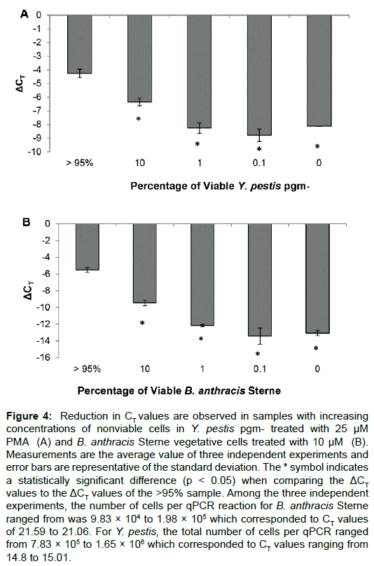
Mixtures of viable and nonviable cells were used in the PMA-qPCR assay to assess the suitability of PMA treatment in situations with mixed samples. All samples contained the same total number of cells, only the ratio of viable to nonviable cells was adjusted. As shown in (Figure 4), >95%, 10%, 1%, 0.1%, or 0% viable cells were mixed with nonviable cells. For Y. pestis pgm- (Figure 4A) and B. anthracis Sterne (Figure 4B), the highest ΔCT values were observed in samples when the majority of the population was comprised of nonviable cells. An analysis workflow depicting the mathematical method for calculating ΔCT values from raw CT values is provided in (Figure 2). The assays for both organisms reached a maximal ΔCT with 1% viable cells, with no significant difference in ΔCT values from the 0.1% or 0% samples (p > 0.05). However, the ΔCT values were significant (p < 0.05) at every tested concentration when compared to the sample with >95% viable cells. The optimized conditions for the PMA assay with the two organisms tested are presented in (Table 3).
Figure 4: Reduction in CT values are observed in samples with increasing concentrations of nonviable cells in Y. pestis pgm- treated with 25 μM PMA (A) and B. anthracis Sterne vegetative cells treated with 10 μM (B). Measurements are the average value of three independent experiments and error bars are representative of the standard deviation. The * symbol indicates a statistically significant difference (p < 0.05) when comparing the ΔCT values to the ΔCT values of the >95% sample. Among the three independent experiments, the number of cells per qPCR reaction for B. anthracis Sterne ranged from was 9.83 × 10⁴ to 1.98 × 10⁵ which corresponded to CT values of 21.59 to 21.06. For Y. pestis, the total number of cells per qPCR ranged from 7.83 × 10⁵ to 1.65 × 10⁶ which corresponded to CT values ranging from 14.8 to 15.01.
Discerning viable from nonviable cells in the presence of white powders.
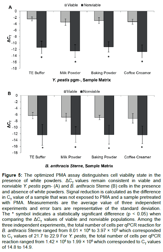
Because intentional release of anthrax spores and stabilized Y. pestis could occur in the form of a powder [28], we tested whether or not the PMA assay was compatible with white powders in the sample matrix. The same criterion of ΔCT value “cut-offs” (refer to equation 1) was used to determine if the white powders interfered with the assay. Using optimized assay conditions (Table 3), we observed ΔCT values of at least six for both Y. pestis pgm- and B. anthracis Sterne cells in assay buffer (Figure 3). If similar magnitudes of the ΔCT value was obtained in samples containing white powders versus samples in buffer alone (i.e., ΔCT value is > 6), we concluded that there was no interfering effect.
We selected milk powder, baking powder, and coffee creamer in powder form to test the effects of the presence of these powders in the samples at a 0.1 mg/mL concentration. As shown in (Figure 5), the ΔCT values in both viable and nonviable samples are consistent in the presence and absence of white powders in comparison to samples that did not contain powders. Comparing the ΔCT values of viable cells to the ΔCT values for nonviable cells was consistently a magnitude of six or higher for both Y. pestis pgm- (Figure 5A) and B. anthracis Sterne vegetative cells (Figure 5B), thereby meeting the criteria for simultaneous identification and viability assessment. The data presented in the figures are based on three independent experiments averaged together. For each independent experiment, the ΔCT (i.e., ΔCTnonviable – ΔCTviable) value was at least 6. However, the ΔCT values in the three independent experiments ranged from six to 15, thereby resulting in a high standard deviation for this data set. From these data, we determined that this assay is effective even in the presence of the tested white powders.
Figure 5: The optimized PMA assay distinguishes cell viability state in the presence of white powders. ΔCT values remain consistent in viable and nonviable Y. pestis pgm- (A) and B. anthracis Sterne (B) cells in the presence and absence of white powders. Signal reduction is calculated as the difference in CT value of a sample that was not exposed to PMA and a sample pretreated with PMA. Measurements are the average value of three independent experiments and error bars are representative of the standard deviation. The * symbol indicates a statistically significant difference (p < 0.05) when comparing the ΔCT values of viable and nonviable populations. Among the three independent experiments, the total number of cells per qPCR reaction for B. anthracis Sterne ranged from 8.01 × 10⁴ to 3.97 × 10⁵ which corresponded to CT values of 21.7 to 22.9 For Y. pestis, the total number of cells per qPCR reaction ranged from 1.42 × 10⁶ to 1.99 × 10⁶ which corresponded to CT values of 14.8 to 14.9.
Discussion
Developing rapid methods for the detection, quantification, and viability determination of B. anthracis and Y. pestis is relevant to several fields of study, including use in forming decisions about the severity of a bio threat event or the safety of food [7,9,12,18,29]. Two drivers for developing a rapid method that simultaneously identifies bacterial species and determines cell viability are related to situations where there is an environmental exposure or identification of a suspicious white powder. First responders need to know whether the sample contains a biological agent, if the agent is a bio threat, and if so, its viability status. The second driver for a rapid viability detection method is in verifying decontamination of areas such as individual rooms of a building after a bio threat event. After decontamination, one could expect nucleic acid or protein fragments to persist, making it possible for qPCR or immunoassays to yield a positive result; however, the result would most likely be a false positive because the biological material would be nonviable and would not pose a threat to human health. In this case, traditional time consuming methods such as cultivation in a laboratory (which takes 24 hours or more) would be necessary prior to detection.
In this study, we tested the suitability of PMA-qPCR to simultaneously identify and assess the viability of B. anthracis Sterne and Y. pestis pgm- without DNA extraction and in the presence of white powders. PMA selectively enters nonviable cells, or cells with damaged membranes, and forms covalent bonds with DNA after photoactivation. PMA assays have been developed to assess the viability of food-associated bacterial pathogens including L. monocytogenes and E. coli O157:H7 [14,30]; however, to the best of our knowledge, this is the first report of a PMA assay for assessing the viability of the bio threat agents B. anthracis and Y. pestis.
Suitable conditions for PMA treatment and detection of viable and non-viable B. anthracis Sterne and Y. pestis pgm- cells are summarized in (Table 3). We explored PMA concentration (Figure 3), LED exposure time, and cellular fitness levels for Y. pestis pgm- and B. anthracis Sterne (data not shown). With respect to cellular fitness, inconsistent results were observed when using overnight cultures that had reached the plateau phase of growth; we identified that consistent results were obtained when cells were in log phase of growth. The consistent change in ΔCT values between viable and nonviable populations was at least two fold under these conditions. Unfortunately, the method was not suitable for use with B. anthracis Sterne spores because viable spores could not be discerned from nonviable spores (see further discussion below). Therefore, the assay is only suitable with cells in a vegetative (i.e., metabolically active) form. Of particular note is that in the more recent of the aforementioned studies [12,31,32], a DNA extraction step was used after PMA treatment to generate the nucleic acid template for the qPCR reaction. In this study, we showed that DNA extraction is not required for obtaining consistent results with the PMA-qPCR assay. The changes in CT values induced by the PMA treatment were similar to those observed by other groups using DNA extracted from samples as the qPCR template rather than intact cells [12,16,33]. These data further suggest that sufficient lysis is occurring during the qPCR reaction to enable access to the DNA for amplification of viable and nonviable cells, thereby eliminating the requirement for a DNA extraction prior to the detection step. Removing the DNA extraction requirement is a significant time reduction in the methodology, especially with respect to Gram-positive bacteria which typically require up to three hours for DNA extraction. Reduced sample processing for metabolically active (i.e., log phase) cells in this case improves assay reliability and accuracy while reducing labor requirements, cost, and length of time of the assay.
Our findings are consistent with other groups that have used PMAqPCR in a laboratory setting, with different organisms, demonstrating that nonviable samples had a high CT shift compared to the equivalent biomass (e.g., CFU) of viable samples after PMA treatment [12,31,32]. As was the case with work done by other groups, the PMA-qPCR assay required evaluation of several variables of the assay for each species used in the study. Specifically, we evaluated the following variables: (1) PMA treatment concentration to ensure a consistent two fold change in CT values between viable and nonviable samples; (2) biomass (cell number) in each sample; and (3) assay buffer [12,14,16,21,31-33]. From our studies, we also concluded that a PMA-qPCR assay can effectively discern both organism identity and viability in a mixed sample of viable and nonviable cells, with the ΔCT value increasing as the percentage of nonviable cells increased. We found that for both organisms, the maximal shift in CT value was obtained in samples in which only 1% of the cells were viable. The assay is sufficiently robust to discern the presence of viable cells in an unknown sample, with the caveat that at least 5 × 10³ cells are present in the qPCR reaction.
With respect to evaluation of the PMA treatment concentration, our study is in agreement with the study conducted by Seinige et al. with C. jejuni which focused on artificially contaminating and then recovering bacteria from chicken leg quarters. They observed that with Campylobacter species, the extent of the ΔCT shift was dependent upon PMA concentration (i.e., increasing PMA concentration increased ΔCT shifts) even among viable cell populations, indicating that at sufficiently high concentrations, PMA is able to penetrate viable cells [12]. We also observed this type of CT shift in viable Y. pestis pgm- (Table 2 and Figure 3A), but did not observe this with vegetative B. anthracis Sterne (Table 1 and Figure 3B). Other bacterial species have similar detection profiles as Y. pestis pgm-, such as L. monocytogenes and S. aureus whereby changes in CT can occur at higher PMA treatment concentrations in viable samples (e.g., >30 μM PMA, data not shown) [14]. In regard to total biomass in the samples used with PMA treatment, we did not observe any significant changes in assay performance between samples containing 105 up to 108 nonviable cells, however, studies conducted with Campylobacter species showed significant changes in assay results in samples containing > 104 nonviable cells [12]. The differences between our study and the Campylobacter study highlights the need for assay optimization at the level of the species. In this study, we have provided conditions for effective PMA-qPCR viability assay of both Y. pestis pgm- and B. anthracis Sterne (Table 3).
Common background matrices that have been used in conjunction with the PMA-qPCR assay include human fecal samples with Bacteroides [32], raw surface water (untreated river water) and sewage waters with Cryptosporidium [31], and recovered solutions from food surfaces with Campylobacter [12]. In these previous studies, the application of the PMA-qPCR assay with more complex matrices resulted in difficulties with reproducibility or loss of signal. Matrices generated from recovered food surfaces have been shown to be less problematic in interfering with assay performance compared to wastewaters [34]. Our study indicates that coffee creamer, milk powder, and baking powder do not interfere with the PMA-qPCR viability assay performance (Figure 5). This is a promising result and indicates that the PMA-qPCR assay could be used for screening suspicious white powders. Given that our results show that detection is possible in a complex matrix (i.e., additions of powders to buffers), it is conceivable that further optimization could enable detection of these organisms in biological fluids and recovered rinses from suspected contaminated meat.
The B. anthracis lifecycle includes a spore form that is in a dormant state which gives rise to the vegetative cell form. Vegetative cells are metabolically active and, upon gaining access to the host, can produce the toxins causing anthrax infection. The spore form has been proven to be robust and resilient to molecular analysis, highlighting the need for developing a rapid viability assay. Although the presence of whole spores can be detected by qPCR using the tested primer/probe set (Table 1), the qPCR assay coupled with PMA treatment does not provide meaningful information with respect to the viability of the spore (Table 1). Although Rawsthorne et al. demonstrated a CT shift in Bacillus subtilis endospores when subjected to PMA treatment [35], we were not able to replicate these results with B. anthracis Sterne spores. We believe there are several reasons for the discrepancy. The first reason is related to the concentration of spores used in our study, which is a relatively small amount of biomass (~15,000 spores). The raw CT values for DNA extracted from viable spores and spores inactivated with hydrogen peroxide were either not detected or greater than 35 (data not shown). We also attempted autoclaving 109 spores and then extracting DNA as was done by Rawsthorne [35], but the extracted DNA from the inactivated samples could not be detected by qPCR either (data not shown). This result is in agreement with the study conducted by Dang et al. which showed that B. anthracis Ames spores that were inactivated by autoclaving had significant decreases in signal in qPCR assays [36]. The second issue in using spores with the PMA-qPCR assay is that the assay is dependent upon a significant shift in CT values between samples treated or untreated with PMA. Because the CT values of DNA extracted from untreated spores was so high, any shift in CT would result in an absence of qPCR signal, resulting in a false negative for identification, without which, a viability assessment cannot be conducted. Because spores were incompatible with the PMA assay, a germination step is required to obtain vegetative cells which are compatible with the developed assay.
In conclusion, we optimized a PMA-qPCR assay for rapid identification and viability analysis of Y. pestis and B. anthracis Sterne vegetative cells, and demonstrated that three different types of white powders do not interfere with the PMA-qPCR assay when conducted in a compatible buffer. However, the assay produces false negative results (i.e., not detected by qPCR) with viable B. anthracis spores, and must be conducted with vegetative cells. Therefore, more research is required to overcome the limitations caused by the required germination step to produce vegetative cells in order to use this rapid viability assay for spore identification and viability analysis.
Conclusions
The developed assay enables simultaneous identification and viability assessment for B. anthracis Sterne and Y. pestis pgm- under laboratory conditions, even in the presence of white powders. Eliminating the DNA extraction step that is typically used reduces total assay time and labor requirements for sample analysis.
Acknowledgements
The research described in this paper was conducted under the Laboratory Directed Research and Development Program at Pacific Northwest National Laboratory, a multiprogramming national laboratory operated by Battelle for the U.S. Department of Energy. Battelle Memorial Institute operates Pacific Northwest National Laboratory for the U.S. DOE under Contract DE-AC06-76RLO.
References
- Mock M, Fouet A (2001) Anthrax. Annu Rev Microbiol 55: 647-671.
- Bartlett JG, Inglesby TV, Borio L (2002) Management of anthrax. Clin Infect Dis 35: 851-858.
- Stewart A, Satterfield B, Cohen M, O'Neill K, Robison R (2008) A quadruplex real-time PCR assay for the detection of Yersinia pestis and its plasmids. Journal of Medical Microbiology 57: 324-331.
- Welch TJ, Fricke WF, McDermott PF, White DG, Rosso M-L, et al. (2007) Multiple Antimicrobial Resistance in Plague: An Emerging Public Health Risk. PLoS ONE 2: e309.
- Gilbert SE, Rose LJ, Howard M, Bradley MD, Shah S, et al. (2014) Evaluation of swabs and transport media for the recovery of Yersinia pestis. J Microbiol Methods 96: 35-41.
- Smith H, Keppie J, Stanley J (1955) The chemical basis of the virulence of Bacillus anthracis. Br J Exp Pathol 36: 460.
- Irenge LM, Gala JL (2012) Rapid detection methods for Bacillus anthracis in environmental samples: a review. Applied Microbiology and Biotechnology 93: 1411-1422.
- Delgado-Viscogliosi P, Solignac L, Delattre JM (2009) Viability PCR, a Culture-Independent Method for Rapid and Selective Quantification of Viable Legionella pneumophila Cells in Environmental Water Samples. Applied and Environmental Microbiology 75: 3502-3512.
- Chen S, Wang F, Beaulieu JC, Stein RE, Ge B (2011) Rapid Detection of Viable Salmonellae in Produce by Coupling Propidium Monoazide with Loop-Mediated Isothermal Amplification. Applied and Environmental Microbiology 77: 4008-4016.
- Taskin B, Gozen AG, Duran M (2011) Selective Quantification of Viable Escherichia coli Bacteria in Biosolids by Quantitative PCR with Propidium Monoazide Modification. Applied and Environmental Microbiology 77: 4329-4335.
- Li B, Chen JQ (2012) Real-Time PCR Methodology for Selective Detection of Viable Escherichia coli O157:H7 Cells by Targeting Z3276 as a Genetic Marker. Applied and Environmental Microbiology 78: 5297-5304.
- Seinige D, Krischek C, Klein G, Kehrenberg C (2014) Co mparative Analysis and Limitations of Ethidium Monoazide and Propidium Monoazide Treatments for the Differentiation of Viable and Nonviable Campylobacter Cells. Applied and Environmental Microbiology 80: 2186-2192.
- Nocker A, Sossa-Fernandez P, Burr MD, Camper AK (2007) Use of Propidium Monoazide for Live/Dead Distinction in Microbial Ecology. Applied and Environmental Microbiology 73: 5111-5117.
- Nocker A, Cheung C-Y, Camper AK (2006) Comparison of propidium monoazide with ethidium monoazide for differentiation of live vs. dead bacteria by selective removal of DNA from dead cells. Journal of Microbiological Methods 67: 310-320.
- Tavernier S, Coenye T (2015) Quantification of Pseudomonas aeruginosa in multispecies biofilms using PMA-qPCR. PeerJ 3: e787.
- Rudi K, Moen B, Drømtorp SM, Holck AL (2005) Use of Ethidium Monoazide and PCR in Combination for Quantification of Viable and Dead Cells in Complex Samples. Applied and Environmental Microbiology 71: 1018-1024.
- Inglis GD, McAllister TA, Larney FJ, Topp E (2010) Prolonged Survival of Campylobacter Species in Bovine Manure Compost. Applied and Environmental Microbiology 76: 1110-1119.
- Chang B, Sugiyama K, Taguri T, Amemura-Maekawa J, Kura F, et al. (2009) Specific Detection of Viable Legionella Cells by Combined Use of Photoactivated Ethidium Monoazide and PCR/Real-Time PCR. Applied and Environmental Microbiology 75: 147-153.
- Cenciarini-Borde C, Courtois S, La Scola B (2009) Nucleic acids as viability markers for bacteria detection using molecular tools. Future Microbiol 4: 45-64.
- Flekna G, Štefanic P, Wagner M, Smulders FJM, Možina SS, et al. (2007) Insufficient differentiation of live and dead Campylobacter jejuni and Listeria monocytogenes cells by ethidium monoazide (EMA) compromises EMA/real-time PCR. Research in Microbiology 158: 405-412.
- Nocker A, Sossa KE, Camper AK (2007) Molecular monitoring of disinfection efficacy using propidium monoazide in combination with quantitative PCR. Journal of Microbiological Methods 70: 252-260.
- Buhr TL, McPherson DC, Gutting BW (2008) Analysis of broth-cultured Bacillus atrophaeus and Bacillus cereus spores. Journal of Applied Microbiology 105: 1604-1613.
- Wielinga PR, Hamidjaja RA, Ågren J, Knutsson R, Segerman B, et al. (2011) A multiplex real-time PCR for identifying and differentiating B. anthracis virulent types. International Journal of Food Microbiology 145, Supplement 1: S137-S144.
- Huang J, Zhu Y, Wen H, Zhang J, Huang S, et al. (2009) Quadruplex Real-Time PCR Assay for Detection and Identification of Vibrio cholerae O1 and O139 Strains and Determination of Their Toxigenic Potential. Applied and Environmental Microbiology 75: 6981-6985.
- Warth A (1978) Molecular structure of the bacterial spore. Adv Microbiol Physiol 17: 1.
- Corfe B, Sammons R, Smith D, Mauël C (1994) The gerB region of the Bacillus subtilis 168 chromosome encodes a homologue of the gerA spore germination operon. Microbiology 140: 471.
- Dragon D, Rennie R (1995) The ecology of anthrax spores: tough but not invincible. Can Vet J 36: 295.
- Parascandola R (2014) Corn starch suspected in white powder scares in at least five hotels near Super Bowl site. New York Daily News. New York, New York.
- Botteldoorn N, Van Coillie E, Piessens V, Rasschaert G, Debruyne L, et al. (2008) Quantification of Campylobacter spp. in chicken carcass rinse by real-time PCR. Journal of Applied Microbiology 105: 1909-1918.
- Pan Y, Breidt F (2007) Enumeration of Viable Listeria monocytogenes Cells by Real-Time PCR with Propidium Monoazide and Ethidium Monoazide in the Presence of Dead Cells. Applied and Environmental Microbiology 73: 8028-8031.
- Brescia CC, Griffin SM, Ware MW, Varughese EA, Egorov AI, et al. (2009) Cryptosporidium propidium monoazide-PCR, a molecular biology-based technique for genotyping of viable Cryptosporidium oocysts. Appl Environ Microbiol 75: 6856-6863.
- Bae S, Wuertz S (2009) Discrimination of Viable and Dead Fecal Bacteroidales Bacteria by Quantitative PCR with Propidium Monoazide. Applied and Environmental Microbiology 75: 2940-2944.
- Wahman DG, Wulfeck-Kleier KA, Pressman JG (2009) Monochloramine Disinfection Kinetics of Nitrosomonas europaea by Propidium Monoazide Quantitative PCR and Live/Dead BacLight Methods. Applied and Environmental Microbiology 75: 5555-5562.
- Varma M, Field R, Stinson M, Rukovets B, Wymer L, et al. (2009) Quantitative real-time PCR analysis of total and propidium monoazide-resistant fecal indicator bacteria in wastewater. Water Research 43: 4790-4801.
- Rawsthorne H, Dock CN, Jaykus LA (2009) PCR-Based Method Using Propidium Monoazide To Distinguish Viable from Nonviable Bacillus subtilis Spores. Applied and Environmental Microbiology 75: 2936-2939.
- Dang JL, Heroux K, Kearney J, Arasteh A, Gostomski M, et al. (2001) Bacillus Spore Inactivation Methods Affect Detection Assays. Applied and Environmental Microbiology 67: 3665-3670.
Relevant Topics
- Anthrax Bioterrorism
- Bio surveilliance
- Biodefense
- Biohazards
- Biological Preparedness
- Biological Warfare
- Biological weapons
- Biorisk
- Bioterrorism
- Bioterrorism Agents
- Biothreat Agents
- Disease surveillance
- Emerging infectious disease
- Epidemiology of Breast Cancer
- Information Security
- Mass Prophylaxis
- Nuclear Terrorism
- Probabilistic risk assessment
- United States biological defense program
- Vaccines
Recommended Journals
Article Tools
Article Usage
- Total views: 11059
- [From(publication date):
August-2015 - Aug 16, 2025] - Breakdown by view type
- HTML page views : 10133
- PDF downloads : 926
