Case Report Open Access
Leiomyosarcoma of the Renal Pelvis: Report of a Case and Review of the Literature
| Een Young Cho1, Jung-Hee Yoon1* and Woogyeong Kim2 | |
| 1Department of Radiology, Inje University Haeundae Paik Hospital, College of Medicine, 1435 Jwa-dong, Haeundae-gu, Busan, 612-030, Republic of Korea | |
| 2Department of Pathology, Inje University Haeundae Paik Hospital, College of Medicine, 1435 Jwa-dong, Haeundae-gu, Busan, 612-030, Republic of Korea | |
| Corresponding Author : | Jung-Hee Yoon Department of Radiology, Inje University Haeundae Paik Hospital, College of Medicine 1435 Jwa-dong, Haeundae-gu, Busan 612-030,South Korea Tel: 82-51-797-0355 Fax: 82-51-797-0379 E-mail: radyjh@hanmail.net |
| Received October 22, 2013; Accepted November 20, 2013; Published November 30, 2013 | |
| Citation: Cho EY, Yoon JH, Kim W (2013) Leiomyosarcoma of the Renal Pelvis: Report of a Case and Review of the Literature. OMICS J Radiology 3:154. doi: 10.4172/2167-7964.1000154 | |
| Copyright: © 2013 Cho EY, et al. This is an open-access article distributed under the terms of the Creative Commons Attribution License, which permits unrestricted use, distribution, and reproduction in any medium, provided the original author and source are credited. | |
Visit for more related articles at Journal of Radiology
Abstract
We report a case of leiomyosarcoma of the renal pelvis developed in 56-year-old man whom underwent MDCT scan presented with hematuria and left flank pain. There was a 7.8×7.6 cm heterogeneous enhanced mass filling in the left renal pelvis with necrotic portion and multiple dystrophic calcifications. The tumor measured about 6.8×5.5 cm in size composed of atypical spindle cells arising from the muscularis mucosa of the renal pelvis. The tumor cells were positive for smooth muscle actin, focal positive for desmin and myoglobin but negative for CD 117 (c-KIT), pancytokeratin and CD34. The histopathologic diagnosis was made as a leiomyosarcoma of the renal pelvis.
| Keywords |
| Leiomyosarcoma; Kidney tumor; Renal pelvic tumor; Renal mass |
| Abbreviation |
| MDCT: Multi-Detector Computed Tomography; HPF: High-Power Fields; SMA: Smooth Muscle Actin; RCC: Renal Cell Carcinoma; TCC: Transitional Cell Carcinoma |
| Introduction |
| Leiomyosarcoma of the renal pelvis is an extremely rare mesenchymal tumor of the kidney. To the best of our knowledge, there is no article that focused on radiologic and pathologic correlation of the renal pelvic leiomyosarcoma. |
| Primary renal sarcomas are rare tumors presenting for 1%-3% of the malignant renal tumors. Leiomyosarcoma is the most common subtype of the renal sarcomas accounting for 50%-60% of all renal sarcomas [1]. Most renal leiomyosarcoma arising from the renal capsule and from smooth muscle of the renal pelvis is uncommon [2]. |
| Here, we describe a rare case of leiomyosarcoma of the renal pelvis detected on the Multi-Detector Computed Tomography (MDCT) with pathologic correlation and we review the previous literature. |
| Case Report |
| A 56-year-old man with hematuria and left flank pain presented to the Inje University Haeundae Paik Hospital. The patient had an open ureterolithotomy for left renal stone 26 years ago and extracorporeal shock wave lithotripsy for same reason 23 years ago. Laboratory data showed no impairment of renal function (blood urea nitrogen and creatinine levels, 10.4 and 1.11 mg/dL, respectively). Routine urinalysis displayed proteinuria (+3) and hematuria (+3). A MDCT scan was performed for further evaluation. MDCT included an unenhanced phase scan, a nephrographic phase scan, a pyelographic phase scan and three-dimensional reformations with coronal and sagittal maximum intensity projections of the kidney. |
| Nephrographic and pyelographic phase scans that were followed with a 100 sec delay after attenuation of the aorta at the thoracolumbar junction had reached 100 HU and a fixed 12 minute delay, respectively, after the intravenous injection of 110 mL of iobitridol (Xenetics; Guerbet, Paris, France) administered at a rate of 3 mL/s with an autonomic injector. |
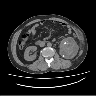
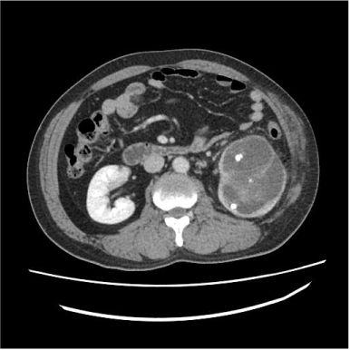
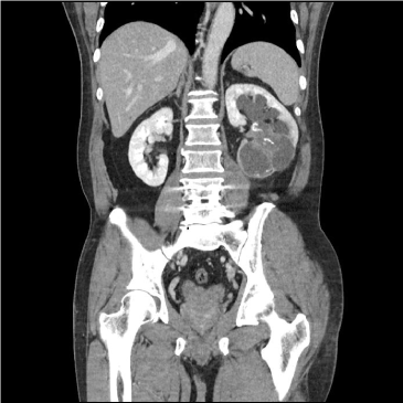
| MDCT scan demonstrated a 7× 6 cm solid and cystic mass with calcifications filling in the left renal pelvis resulting in hydronephrosis but preserved the reniform. The enhancement pattern is heterogeneous and gradual; diffuse gradual enhancement and some portions showed relatively low attenuation (Figures 1A-1C). Peripheral to the solid portion of the tumor cystic portions was present, intracystic attenuation showed heterogeneous and high attenuation than that of dilated renal calices (Figure 1D). Multiple calcifications of variable size and shape were at both solid component and cystic component. The tumor located in renal pelvic and inferior portion of the kidney, thinning of renal cortex and focal obliteration of thinned renal cortex with perinephric fat infiltration was noted. There was no mass at contralateral kidney, both ureters and urinary bladder. There was no enlarged lymph node at the regional or distant area. Preoperative differential diagnosis was urothelial cell carcinoma such as transitional cell carcinoma, squamous cell carcinoma or mucinous adenocarcinoma considering prevalence and patient’s history. |
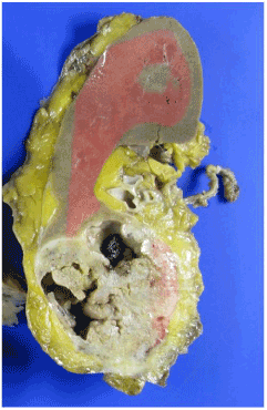
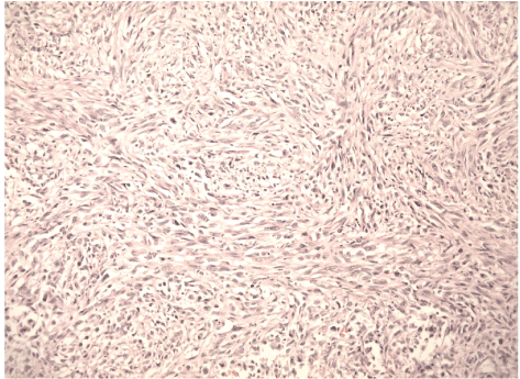
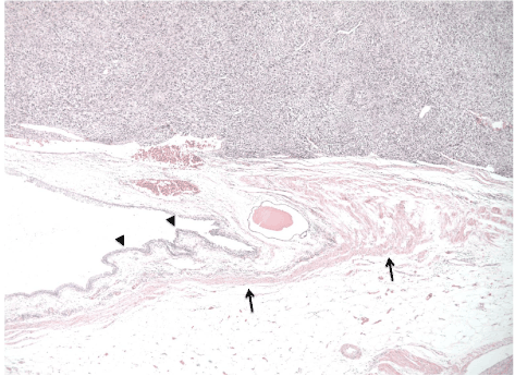
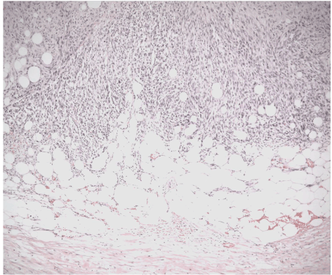
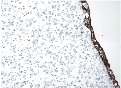
| Laparoscopic left nephroureterectomy was performed. Grossly the tumor was measured 6.8×5.5 cm identified in lower portion of the kidney (Figure 2A). The cut surface was grayish white solid. Microscopically, the tumor was composed of atypical spindle cells that were arranged as alternating fascicles (Figure 2B). The mass originating from the muscularis mucosa layer of renal pelvis contained dystrophic calcifications and necrosis with invasion of renal parenchyma and perinephric fat tissue (Figures 2C and 2D) with underlying chronic pyelonephritis. The number of mitotic activity was 19 mitoses per 10 High-Power Fields (HPF) on average. Immunohistochemically, the tumor cells were positive for Smooth Muscle Actin (SMA) (Figure 3A), and negative for pancytokeratin thus sarcomatoid carcinoma could be ruled out (Figure 3B). Additional immune histochemistry for vimentin was positive, desmin and myoglobin were positive and CD 34 and EMA were negative. Based on the histopathology and immunohistochemistry, the mass was diagnosed as leiomyosarcoma arising from the renal pelvis. A pulmonary nodule was newly developed on 2 months follow up chest CT and PET CT. |
| Discussion |
| Leiomyosarcoma from the renal pelvis is a rare disease entity. 95% of the malignant renal pelvic masses are urothelial cell origin tumor. Only 1-3% accounts for renal sarcomas, and leiomyosarcoma is the most common sarcoma of the kidney accounting for 50-60% of cases. Renal leiomyosarcoma mostly occur in adults and no male and female are equally affected. Symptoms are non-specific; include flank pain, palpable mass and hematuria not much different from other renal tumors. Usually metastasize to lung, liver and bone [3]. Leiomyosarcoma may arise from the renal capsule, renal parenchyma, pelvic muscularis, or the main renal vein [3]. Macroscopically, large solid grey-white, soft to firm mass and it may have focally necrotic areas [3]. Renal leiomyosarcoma have no pathognomonic finding that lead to make preoperative diagnosis. In most of the cases, diagnosis of renal leiomyosarcoma would be made after surgical resection and histopathologic examination. |
| The Table summarizes the clinico-radiological features of the previously reported cases in English literature including the present case. To date, there is no sex predilection, and occurred at wide range of age (23-65 years, mean; 47 years). Renal pelvic leiomyosarcoma ranges in size from 2 to 10 cm with a mean of 5.7 cm. A common characteristic feature is filling defect of renal pelvis on retrograde pyelography or intravenous pyelography (Table 1) and solid mass filling in the renal pelvis on contrast enhanced CT (Table 1). This means intraluminal growth pattern of the tumor. Image findings and histopathologic specimen showed intraluminal growth pattern of the tumor more than outward growth pattern. But the tumor infiltrates to perirenal adipose tissue and renal sinus as well as renal parenchyma. Among 6 cases performed contrast enhanced CT, poor (Table 1) and heterogeneous (Table 1) enhancement patterns were shown after contrast administration. Only one case had reported focal necrosis of the tumor (Table 1) as in our case. Furthermore, none of the case reported intratumoral dystrophic calcifications which were found in our case. Our pathologist suggested for dystrophic calcifications rather than renal calculi. There was no reported lymph node metastasis in all cases. |
| Renal leiomyosarcoma was known to have poor prognosis, with a 5 year survival rate of 29-36%. However, in reported renal pelvic leiomyosarcoma cases, the follow-up duration was too short and the number of reported cases is small to precisely predict the prognosis. |
| In 5 cases, preoperative radiologic diagnosis was urothelial cell carcinoma (Table 1) or Renal Cell Carcinoma (RCC) (Table 1). Possible cause of radiologic misdiagnosis is the low prevalence rate of renal pelvic leiomyosarcoma. |
| The majority malignant tumor arising from renal pelvis is urothelial cell carcinoma including Transitional Cell Carcinoma (TCC), squamous cell carcinoma and mucinous adenocarcinoma. Among them TCC is the most common urothelial neoplasm hematuria as common clinical presentation and well known as multiplicity. TCC is papillary intraluminal mass, spreads centrifugally and dilates collecting system [12]. Thus, hydrocalices, amputated calices or phantom calices are shown on excretory urography. On CT scan after contrast injection the mass typically enhances, but lesser degree than normal renal parenchyma [12]. TCC could be noncalcified and calcified. Squamous cell carcinomas and mucinous adenocarcinomas are strongly related with renal calculi and hard to distinguish from TCC [13]. |
| RCCs projecting into renal sinus also should be considered, which also can lead to hydronephrosis or caliceal displacement. However, well enhancement on post contrast CT images will be shown unlike TCCs. In histopathological examination the most important disease to differentiate from leiomyosarcoma is sarcomatoid RCC. Negative for Pancytokeratin immunochemical stain can rule out sarcomatoid RCC. |
| In conclusion, we report a case of leiomyosarcoma arising from renal pelvis, rarely reported malignant mesenchymal neoplasm of the kidney. CT scan of this rare tumor showed heterogeneously enhancing large mass filling in the renal pelvis with cystic change and calcifications. Leiomyosarcomas have histological characterization of alternating fascicular haphazard pattern, bland spindle cells and nuclear atypia, pleomorphism, and necrosis. They confirmed by immunohistochemistry, the tumor cells of leiomyosarcoma are positive for SMA and desmin and the tumor cells are negative for pancytokeratin, CD 117 (c-KIT) and CD34. These histological and immunohistochemical features are helpful in diagnosing this uncommon disease. Our report may help increase the awareness of this rare disease entity and take account of differential diagnosis for renal pelvic mass. |
| Authors Contribution |
| EY Jo: Clinical study, manuscript editing; JH Yoon: Literature search, manuscript editing; W Kim: Pathologic editing. |
References |
|
--
Tables and Figures at a glance
| Table 1 |
Figures at a glance
 |
 |
 |
 |
 |
| Figure 1a | Figure 1b | Figure 1c | Figure 1d | Figure 2a |
 |
 |
 |
 |
 |
| Figure 2b | Figure 2c | Figure 2d | Figure 3a | Figure 3b |
Relevant Topics
- Abdominal Radiology
- AI in Radiology
- Breast Imaging
- Cardiovascular Radiology
- Chest Radiology
- Clinical Radiology
- CT Imaging
- Diagnostic Radiology
- Emergency Radiology
- Fluoroscopy Radiology
- General Radiology
- Genitourinary Radiology
- Interventional Radiology Techniques
- Mammography
- Minimal Invasive surgery
- Musculoskeletal Radiology
- Neuroradiology
- Neuroradiology Advances
- Oral and Maxillofacial Radiology
- Radiography
- Radiology Imaging
- Surgical Radiology
- Tele Radiology
- Therapeutic Radiology
Recommended Journals
Article Tools
Article Usage
- Total views: 14322
- [From(publication date):
January-2014 - Aug 29, 2025] - Breakdown by view type
- HTML page views : 9672
- PDF downloads : 4650
