Case Report Open Access
Sialolipoma of Minor Salivary Gland in Uvula
Basak K*, Kayipmaz S and Karadayi N
Department of Pathology, Semsi Denizer Cad. E-5 Karayolu, Cevizli Mevkii, Istanbul, Turkey
- *Corresponding Author:
- Kayhan Basak
Dr.Lütfi Kirdar Kartal Education and Research Hospital
Department of Pathology, Semsi Denizer Cad. E-5 Karayolu
Cevizli Mevkii, 34890 Kartal, Istanbul, Turkey
Tel: 90 216 4413900
Fax: 90 216 3520083
E-mail: kayhan.basak@sbkeah.gov.tr
Received Date: August 06, 2014; Accepted Date: August 21, 2014; Published Date: August 26, 2014
Citation: Basak K, Kayipmaz S, Karadayi N (2014) Sialolipoma of Minor Salivary Gland in Uvula. J Oral Hyg Health 2:159. doi:10.4172/2332-0702.1000159
Copyright: © 2014 Basak K, et al. This is an open-access article distributed under the terms of the Creative Commons Attribution License, which permits unrestricted use, distribution, and reproduction in any medium, provided the original author and source are credited.
Visit for more related articles at Journal of Oral Hygiene & Health
Abstract
Lipomatous lesions of the salivary glands are rare, accounting for less than 0.5% of all parotid gland tumours. Distinct microscopic variants of lipoma of the salivary glands, e.g. angiolipoma, fibrolipoma, pleomorphic lipoma and spindle-cell lipoma have been reported. A 45 year-old male patient with mass on uvula was presented. The specimen was capsulated, yellow coloured, soft tissue with 1.3 cm in greatest diameter. Whole-mount section showed tumor composed by mature adipocytes, salivary gland parenchymal tissue and lymphoid follicles surrounded by a fibrous capsule. Salivary gland component consist acinar and ductal elements. In some areas, glandular components were atrophic. Lymphoid follicles and focal fibrosis were seen. Oncocytic, sebaceous, and squamous metaplasia were not observed. Sialolipomas were composed predominantly of adipose tissue and showed expansive growth with fibrous capsule. Sialolipomas were reported at parotid gland, submandibular gland, hard and soft palate. To our knowledge, such a case in uvula localization was not previously presented.
Keywords
Sialolipoma; Minor salivary gland; Uvula
Introduction
Lipomatous lesions of the salivary glands are rare accounting for less than 0.5% of all parotid gland tumours [1]. Although distinct microscopic variants of lipoma of the salivary glands, e.g. angiolipoma, fibrolipoma, pleomorphic lipoma and spindle-cell lipoma have been reported [2-5]. Term of sialolipoma was first used by Nagao et al. [6]. The patients were from birth to 84 years old, and average of age was 55.7 years [6-11]. Male cases were slightly more common than female ones6. Sialolipomawas reported to occur in both major and minor salivary glands [1-5].
Case Report
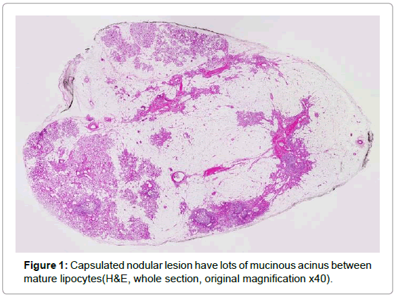
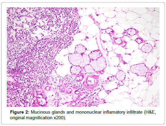
A forty five-year-old male patient was presented with a mass on uvula. The specimen was a capsulated, yellow coloured, soft tissue, 1.3 cm in greatest diameter. Cut surface was solid and yellow. Wholemount section showed that tumor composed by mature adipocytes, salivary gland parenchymal tissue and lymphoid follicles surrounded by a fibrous capsule. Salivary gland component consists acinar and ductal elements (Figure 1). In some areas, glandular components were atrophic. Lymphoid follicles and focal fibrosis were seen (Figure 2). Oncocytic, sebaceous, and squamous metaplasia was not observed.
Discussion
Sialolipomas were predominantly composed of adipose tissue and showed expansive growth with fibrous capsule. Sialolipomas were previously reported at parotid and submandibular glands [11,12] and can occur almost any site other than the salivary glands [6,10]. Qayyum et al. reviewed 35 cases and documented that sialolipoma of minor salivary gland were reported only in adults [10]. The glandular components closely resembled the normal salivary gland parenchyma without any atypia, albeit with the presence of minor metaplastic changes [6]. In our case, metaplastic changes were not observed but contain inflammatory infiltration with lymphoid follicles. Immunohistological and ultrastructural studies confirmed that the glandular components become entrapped during lipomatous proliferation, rather than representing true neoplastic elements [6]. These findings suggested sialolipoma as a distinct variant of salivary gland lipoma.
References
- Ellis GL, Auclair PL (1996)Tumors of the Salivary Glands. Atlas of Tumor Pathology. Washington DC: Armed Forces Institute of Pathology.
- Reilly JS, Kelly DR, Royal SA (1988) Angiolipoma of the parotid: case report and review. Laryngoscope 98: 818-821.
- Hatziotis JC (1971) Lipoma of the oral cavity. Oral Surg Oral Med Oral Pathol 31: 511-524.
- Graham CT, Roberts AH, Padel AF (1998) Pleomorphic lipoma of the parotid gland. J LaryngolOtol 112: 202-203.
- deMoraes M, de Matos FR, de Carvalho CP, de Medeiros AM, de Souza LB (2010) Sialolipoma in minor salivary gland: case report and review of the literature. Head Neck Pathol 4: 249-252.
- Nagao T, Sugano I, Ishida Y, Asoh A, Munakata S, et al. (2001) Sialolipoma: a report of seven cases of a new variant of salivary gland lipoma. Histopathology 38: 30-36.
- Hornigold R, Morgan PR, Pearce A, Gleeson MJ (2005) Congenital sialolipoma of the parotid gland first reported case and review of the literature. Int J PediatrOtorhinolaryngol 69: 429-434.
- Ramer N, Lumerman HS, Ramer Y (2007) Sialolipoma: report of two cases and review of the literature. Oral Surg Oral Med Oral Pathol Oral RadiolEndod 104: 809-813.
- Bansal B, Ramavat AS, Gupta S, Singh S, Sharma A, et al. (2007) Congenitalsialolipoma of parotid gland: a report of rare and recently described entity with review of literature. PediatrDevPathol 10: 244-246.
- Qayyum S, Meacham R, Sebelik M, Zafar N (2013) Sialolipoma of the parotid gland: Case report with literature review comparing major and minor salivary gland sialolipomas. J Oral MaxillofacPathol 17: 95-97.
- Herrera ÓM, Rodríguez RR, Noriega JCL (2013)Surgical excision of sialolipoma. Report of clinical case. RevistaOdontolÓgica Mexicana 17:121-124.
- Agaimy A, Ihrler S, Markl B, Lell M, Zenk J, et al (2013) Lipomatous Salivary Gland Tumors: A Series of 31 Cases Spanning Their Morphologic Spectrum With Emphasis on Sialolipoma and OncocyticLipoadenoma. Am J SurgPathol 37:128- 137.
Relevant Topics
- Advanced Bleeding Gums
- Advanced Receeding Gums
- Bleeding Gums
- Children’s Oral Health
- Coronal Fracture
- Dental Anestheia and Sedation
- Dental Plaque
- Dental Radiology
- Dentistry and Diabetes
- Fluoride Treatments
- Gum Cancer
- Gum Infection
- Occlusal Splint
- Oral and Maxillofacial Pathology
- Oral Hygiene
- Oral Hygiene Blogs
- Oral Hygiene Case Reports
- Oral Hygiene Practice
- Oral Leukoplakia
- Oral Microbiome
- Oral Rehydration
- Oral Surgery Special Issue
- Orthodontistry
- Periodontal Disease Management
- Periodontistry
- Root Canal Treatment
- Tele-Dentistry
Recommended Journals
Article Tools
Article Usage
- Total views: 16257
- [From(publication date):
November-2014 - Aug 15, 2025] - Breakdown by view type
- HTML page views : 11471
- PDF downloads : 4786


