Research Article Open Access
Protective Immunodominant Zaire Ebolavirus Glycoprotein Epitope in Mice
Xiangguo Qiu1, Lisa Fernando1, Steven M. Jones1,2,3,4 and Judie B. Alimonti1,2*
1Special Pathogens Program, National Microbiology Laboratory, Public Health Agency of Canada, 1015 Arlington St. Winnipeg, Manitoba, R3E 3R2, Canada
2Department of Medical Microbiology, University of Manitoba, Winnipeg, Manitoba, R3E 3R2, Canada
3Department of Immunology, University of Manitoba, Winnipeg, Manitoba, R3E 3R2, Canada
4Cognoveritas Consulting Inc. A-137 Westchester Dr., Winnipeg, Manitoba, R3P 2G6 Canada
- *Corresponding Author:
- Judie Alimonti
Special Pathogens Program, National Microbiology Laboratory
Public Health Agency of Canada, 1015 Arlington St.
Winnipeg, Manitoba, R3E 3R2
Tel: 204-784-5998 or 789-5097
Fax: 204- 789-2140
E-mail: judie.alimonti@phac-aspc.gc.ca
Received Date: August 26, 2011; Accepted Date: October 18, 2011; Published Date: October 20, 2011
Citation: Qiu X, Fernando L, Jones SM, Alimonti JB (2011) Protective Immunodominant Zaire Ebolavirus Glycoprotein Epitope in Mice. J Bioterr Biodef S1:006. doi: 10.4172/2157-2526.S1-006
Copyright: © 2011 Qiu X, et al. This is an open-access article distributed under the terms of the Creative Commons Attribution License, which permits unrestricted use, distribution, and reproduction in any medium, provided the original author and source are credited.
Visit for more related articles at Journal of Bioterrorism & Biodefense
Abstract
Zaire Ebolavirus (ZEBOV) causes a highly lethal severe haemorrhagic fever in humans, for which there is currently no approved vaccine available. In this study, cytokine ELISPOT assays were used to determine the ZEBOVGP-specific cytokine profile and immunodominant T cell epitopes in VSV?G/ZEBOVGP immunized Balb/C mice. Splenocytes added to pools of overlapping peptides spanning ZEBOVGP stimulated a Th1 cytokine profile with the majority of the ZEBOVGP-specific splenocytes secreting IFN-g. The splenocytes produced the strongest IFN-g response to the immunodominant peptide HNTPVYKLDISEATQ, located in the cytopathic mucin domain of GP. The immunodominant epitope was then tested for its ability to induce a protective immune response in mice challenged with a lethal dose of mouse adapted-ZEBOV. Mice immunized with the peptide plus Freund’s adjuvant survived the challenge, and had a strong ZEBOVGP-specific T and B cell immune response. Identifying the ZEBOVGP-specific cytokine profile and immunodominant epitope will aid in determining the correlates of immune protection; allow for the development of assays to assess the efficacy of the VSV?G/ZEBOVGP vaccine in immunized individuals; and provide valuable information for the development of a subunit vaccine.
Keywords
Cytokines; Ebola; Epitope; Glycoprotein; IFN-g; Mice; Mouse adapted-Ebola; T cell; Vaccine; VSV
Abbreviations
eVLPs: Ebola Virus Like Particles; i.p.: Intraperitoneally; KLH: Keyhole Limpet Hemocyanin; MA-ZEBOV: Mouse Adapted ZEBOV; NHP: Nonhuman Primate; NP: Nucleoprotein; PA: Peptide without Freund’s adjuvant; PF: Peptide with Freund’s adjuvant; PK: Peptide-KLH conjugate; PKF: Peptide-KLH conjugate with Freund’s adjuvant; VSV: Vesicular Stomatitis Virus; ZEBOV: Zaire ebolavirus
Introduction
Zaire Ebolavirus is a filovirus that causes a severe haemorrhagic fever in humans with a fatality rate of 50-90%. There are currently no vaccines or effective therapies commercially available. However, due to a rapid and high fatality rate there is need for a vaccine. We have generated a vaccine (VSVΔG/ZEBOVGP) that uses a live attenuated recombinant Vesicular Stomatitis Virus (VSV) with the VSV glycoprotein replaced by the glycoprotein (GP) of ZEBOV [1]. In nonhuman primates (NHPs) the VSVΔG/ZEBOVGP vaccine provides 100% protection against a lethal high dose ZEBOV challenge, and induces long lasting humoural and cellular immunity [2,3]. Although antibody and T cell immune responses have been generated against the ZEBOV GP and nucleoprotein (NP), numerous studies have demonstrated that GP is sufficient in conferring protection [4,5]. As the VSVΔG/ZEBOVGP vaccine is effective in NHPs it would be a useful model in which to study the immunological mechanisms of protection. Therefore determining the cytokine profile and immunodominant T cell epitope would be instrumental in the first steps of identifying putative protective mechanisms for ZEBOV, and aid in developing assays to establish the efficacy of the vaccine in eliciting a protective immune response in immunized individuals.
One of the most effective assays for examining the correlates of immune protection is the ELISPOT assay. The ELISPOT assay is a reliable, reproducible assay for the determination of T cell responses after vaccination, and has been used for many clinical trials to evaluate the immunological efficacy of a vaccine [6-8]. The ELISPOT assay, which can quantify low frequency immune responses on a per cell basis, provides information on the frequency (clonal size), and the cytokine effector signature of the Ag-specific T cell pool in vivo for a primed immune response [9]. In this study, peptides spanning the entire region of ZEBOVGP were added to a cytokine ELISPOT assay in order to determine the ZEBOVGP-specific cytokine profile, as well as the immunodominant ZEBOVGP epitope in VSVΔG/ZEBOVGP immunized mice. The immunodominant peptide was then tested in a mouse model for its ability to provide protection to a lethal challenge of mouse adapted-Zaire Ebolavirus (MA-ZEBOV).
Materials and Methods
Animals and ethics
Female Balb/C mice aged 6-8 weeks were obtained from Charles River (Quebec, Canada). The animal studies were approved by the Canadian Science Centre for Human and Animal Health Care Committee following the guidelines of the Canadian Council on Animal Care. The mice were housed in either the Biosafety level laboratory 2 (BSL2) or BSL4 in environmentally enriched sterile housing with food and water ad libitum.
Vaccine and peptides
The recombinant VSVΔG/ZEBOVGP vaccine expressing the glycoprotein (GP) of ZEBOV (strain Kikwit) were generated using VSV (Indiana serotype) as described previously [1,10]. The 167 peptide library (Mimotopes, Australia) were dissolved in 80% Dimethyl sulfoxide (DMSO) and consisted of 15mers with 11 amino acid overlaps, and spanned the entire 676 amino acids of the ZEBOV, strain Kikwit 1995 GP. Initially, three peptide pools were made: pool 1 (peptides #1-56), pool 2 (peptides #57-112), and pool 3 (peptides #113- 167). Later a second batch of peptide pools were generated from pool # 2 (Table 1). The matrix was designed so that each peptide was present in two pools. In order for a response to an individual peptide to be considered positive it had to be positive in both pools. This allowed the identification of individual immunodominant peptides that could then be tested.
| Pool 9 | Pool 10 | Pool 11 | Pool 12 | Pool 13 | Pool 14 | Pool 15 | Pool 16 | |
|---|---|---|---|---|---|---|---|---|
| Pool 1 | 53 | 54 | 55 | 56 | 57 | 58 | 59 | 60 |
| Pool 2 | 61 | 62 | 63 | 64 | 65 | 66 | 67 | 68 |
| Pool 3 | 69 | 70 | 71 | 72 | 73 | 74 | 75 | 76 |
| Pool 4 | 77 | 78 | 79 | 80 | 81 | 82 | 83 | 84 |
| Pool 5 | 85 | 86 | 87 | 88 | 89 | 90 | 91 | 92 |
| Pool 6 | 93 | 94 | 95 | 96 | 97 | 98 | 99 | 100 |
| Pool 7 | 101 | 102 | 103 | 104 | 105 | 106 | 107 | 108 |
| Pool 8 | 109 | 110 | 111 | 112 | 113 | 114 | 115 | 116 |
Table 1: Peptide Pool Matrixa.
Immunization and splenocyte preparation
The mice were acclimatized for 1 week prior to immunization. Mice received 2x10⁴ pfu VSVΔGZEBOVGP intraperitoneally (i.p.) at least one month prior to boosting with a second immunization. At 14 days after the boost, the spleen was homogenized by passing it through a wire mesh in RPMI 1640, strained through a 40 mm filter, then centrifuged at 450xg for 10 minutes, before resupension in RPMI 1640, 10% heat inactivated fetal bovine serum, 100 U penicillin, 100 mg streptomycin, 2 mM L-glutamine, 25 mM Hepes, non-essential amino acids, 1 mM sodium pyruvate, 50 mM β-mercaptoethanol. They were strained again before addition to the ELISPOT assay plates.
ELISPOT assay
Mouse IFN-g, TNF, IL-2, IL-4, and IL-10 BD ELISPOT assays (BD Bioscience) were performed as per manufacturer’s instructions. Briefly, 5x10⁵ splenocytes/well were incubated with 2 mg/ml of each ZEBOV GP peptide. For the positive controls the mitogens 0.5 mg/ml PMA plus 5 ng/ml Ionomycin was used for IFN-g, and IL-2; 1 mg/ml LPS was used for TNF and IL-10; and 5 mg/ml Con A was used for IL-4. For the negative control 80% DMSO was added at an equivalent volume as the peptides added. Spots were acquired by an automated AID ELISPOT Reader System (CTL, Cleveland, OH) using ELISPOT software version 3.5. The ELISPOT response was considered positive when the number of specific spots/million splenocytes was greater than 4 standard deviations above the average of the DMSO control wells. In addition, the spots in the DMSO negative control well had to be below 50. The results shown in the figures are after the DMSO background was subtracted for each mouse.
Murine protection study
Groups of Balb/C mice (n=7) were immunized subcutaneously with media only (medium), 50 mg of peptide (PF) with or without (PA) Freund’s complete adjuvant, or 50 mg of peptide conjugated to Keyhole Limpet Hemocyanin (KLH) with (PKF) or without (PK) Freund’s complete adjuvant. The mice were immunized twice more, 28 days apart, but with Freund’s incomplete adjuvant used in place of Freund’s complete adjuvant. A control group of mice were immunized intraperitoneally with 2 x 10⁴ pfu of VSVΔG/ZEBOVGP (rVSV) 28 days before the challenge. At 28 days after the last immunization all groups of mice (n=5) were challenged intraperitoneally with 1000 LD50 of mouse adapted Zaire Ebolavirus (MA-ZEBOV) [11]. The survival of the challenged mice was followed for 28 days, which is 3 times longer than the length of time to death of the media only group. The splenocytes for the remaining 2 mice from each group were used in an IFN-g and IL-2 ELISPOT assay two weeks after the third immunization. The 2 mice in the positive control group (rVSV) which received only one immunization were re-immunized 28 days later and the splenocytes assayed two weeks later.
Indirect ELISA
The generation of the Ebola virus like particles (eVLPs) containing the ZEBOV GP1,2 strain Mayinga and VP40 was described previously [12,13], and the eVLPS were used as the antigen in the ELISA. There is only 11 amino acids difference between the Mayinga and Kikwit strains, and they are highly cross-reactive in this assay. High binding polysterene microtitre plates (Thermo) were coated with 60 ml of the eVLP diluted at 5 mg/ml in PBS for 1 hour at 37ºC. The wells were washed 4X with PBS, 1% Tween-20 then blocked with PBS, 2% skim milk (blocking solution) overnight at 4ºC. MAbs diluted in blocking solution (150ml) were added and incubated at 37ºC for 1 hour. After washing the plates 4X, the peroxidase-conjugated affinity purified goat-anti-mouse IgG detection antibody (Rockland) diluted 1:2000 in blocking solution was added for 1 hour at 37ºC. After washing 4X, the substrate (ABTS + H2O2, 100 ml/well) was added and the plates incubated at 37ºC. After 30 minutes, the reaction was read at room temperature with a spectrophotometer (Versamax microplate reader) at 405 nm. All samples were tested in triplicate.
Statistics
All statistics were performed using the Graph Pad Prism v4 software. T tests and Kaplan-Meier survival curve statistics compared the test mice to the media control mice.
Results
Determination of the cytokine profile and immunodominant ZEBOVGP epitope in splenocytes from VSVΔG/ZEBOVGP immunized mice.
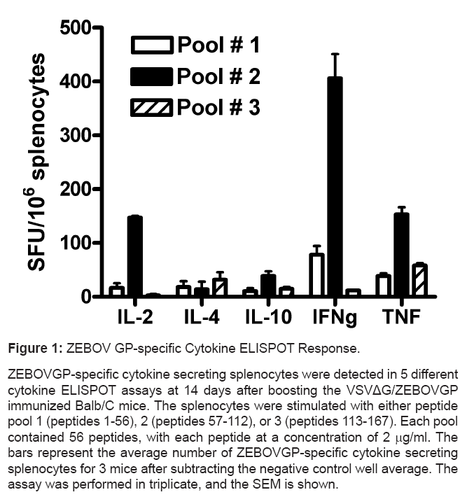
As the ELISPOT assay is a reliable assay for the determination of T cell immune responses to antigens in vaccines, the assay was used to determine what the cytokine profile and immunodominant ZEBOVGP epitope was in mice immunized with the VSVΔG/ZEBOVGP vaccine. Initially, VSVΔG/ZEBOVGP immunized splenocytes from Balb/C mice were added to a range of cytokine ELISPOT assays in order to determine which cytokines were predominantly invoked by the vaccine (Figure 1). For this initial assay 3 pools of 15mer peptides with 11 amino acid overlaps were generated spanning the entire ZEBOVGP. Each pool contained 56 peptides. The VSVΔG/ZEBOVGP immunized splenocytes produced strong signals in pool #2 for the IFN-g, IL2, and TNF ELISPOT assays (Figure 1). However, there was a negative test result for the IL-4 and IL-10 assays as the signal was below the cutoff point for the wells containing the ZEBOVGP peptides. The positive controls for IL-4 and IL-10 worked confirming that the assay worked but the test results were negative. As the IFN-g ELISPOT assay produced the strongest and most reproducible response, this assay was then used to determine the immunodominant epitope.
Figure 1: ZEBOV GP-specific Cytokine ELISPOT Response.
ZEBOVGP-specific cytokine secreting splenocytes were detected in 5 different
cytokine ELISPOT assays at 14 days after boosting the VSVΔG/ZEBOVGP
immunized Balb/C mice. The splenocytes were stimulated with either peptide
pool 1 (peptides 1-56), 2 (peptides 57-112), or 3 (peptides 113-167). Each pool
contained 56 peptides, with each peptide at a concentration of 2 mg/ml. The
bars represent the average number of ZEBOVGP-specific cytokine secreting
splenocytes for 3 mice after subtracting the negative control well average. The
assay was performed in triplicate, and the SEM is shown.
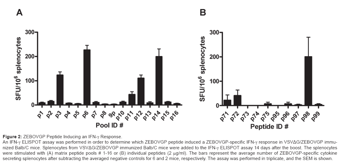
The T cell immunodominant epitope in ZEBOVGP is unknown. To further define the immunodominant epitopes the IFN-g ELISPOT assay was utilized using the peptides from pool # 2. All of the peptides from the second pool plus some neighbouring peptides were assigned to pools, numbered 1-16, based on a matrix configuration (Table 1). While each pool contained 8 peptides, each peptide was present in two pools. In order for a response to be considered positive for a specific peptide the signal had to be positive in both pools. There were very strong signals in pools 3, 6, 12, and 14. Using the matrix, peptides 72, 74, 96, and 98 were identified as possible immunodominant epitopes (Figure 2a). In order to confirm the results, the IFN-g ELISPOT assay was repeated with the individual peptides (Figure 2b). Peptide # 98 produced the strongest signal whereas peptide # 72 produced a much weaker signal, and the remaining neighbouring peptides produced background levels. Therefore the immunodominant epitope is peptide # 98 which contains the following amino acid sequence, HNTPVYKLDISEATQ and is found at amino acids #389-403 of GP.
Figure 2: ZEBOVGP Peptide Inducing an IFN-g Response.
An IFN-g ELISPOT assay was performed in order to determine which ZEBOVGP peptide induced a ZEBOVGP-specific IFN-g response in VSVΔG/ZEBOVGP immunized
Balb/C mice. Splenocytes from VSVΔG/ZEBOVGP immunized Balb/C mice were added to the IFN-g ELISPOT assay 14 days after the boost. The splenocytes
were stimulated with (A) matrix peptide pools # 1-16 or (B) individual peptides (2 mg/ml). The bars represent the average number of ZEBOVGP-specific cytokine
secreting splenocytes after subtracting the averaged negative controls for 6 and 2 mice, respectively. The assay was performed in triplicate, and the SEM is shown.
Murine protection study
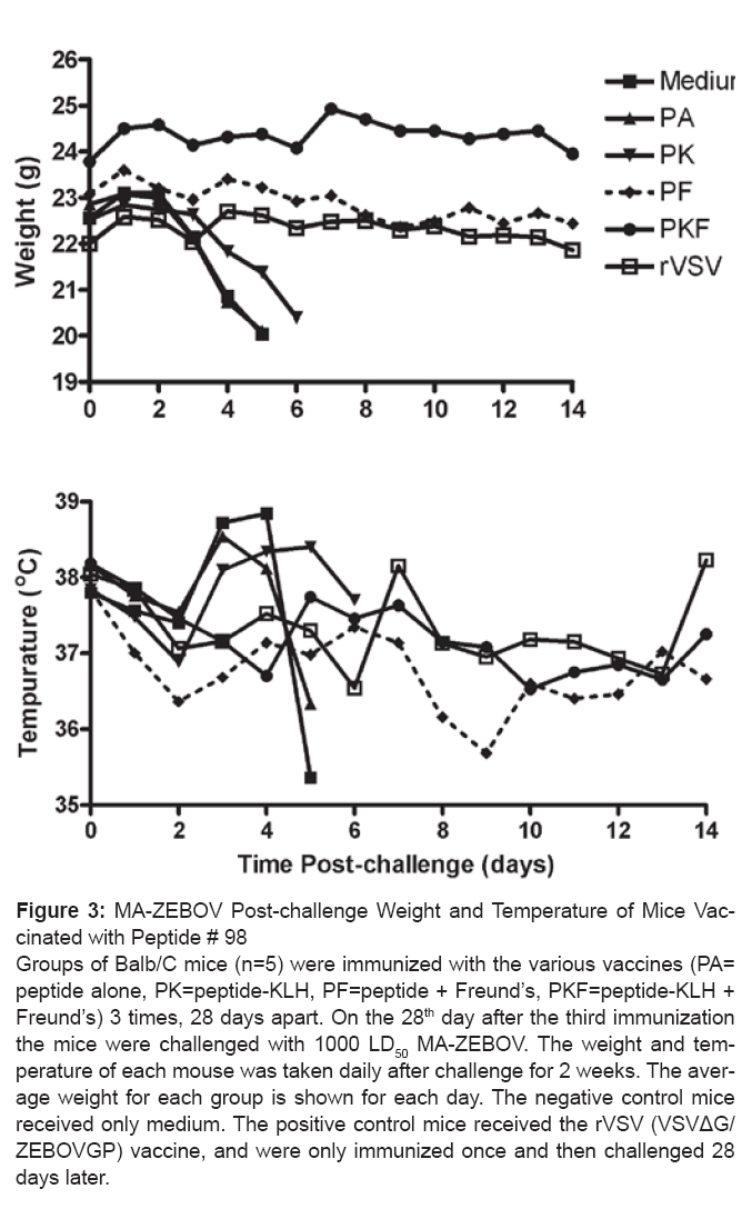
In order to determine whether the immunodominant HNTPVYKLDISEATQ peptide # 98 could induce a protective immune response, the peptide was used to immunize mice before a lethal high dose challenge with MA-ZEBOV (Table 2). As peptides alone generate poor immune responses the peptide was also used in conjunction with the carrier protein KLH, or Freund’s adjuvant. Groups of mice (n=5) were immunized subcutaneously with 50 mg of peptide, 3 times, 28 days apart before being challenged with 1000 LD50 MA-ZEBOV. A media only negative control group was used to demonstrate the lethality of the MA-ZEBOV infection. The VSVΔG/ZEBOVGP (rVSV) vaccine given 28 days prior to challenge was used as a positive control to demonstrate complete protection against a high dose MA-ZEBOV infection [14]. All of the mice in the media only, peptide alone (PA), and peptide-KLH (PK) conjugated groups died by day 8. The mice in all 3 groups showed signs of illness as indicated by a significant drop in weight, along with an increased temperature on day 3 and 4 which decreased below normal shortly thereafter (Figure 3). However, while there was no difference in the mean time to death between the media and PA groups (5.7 and 5.5 days, respectively), the mice immunized with the PK had a mean time to death of 6.5 days which was significantly longer (p=0.016) than the media only group. The two groups of mice that were immunized with the peptide plus Freund’s adjuvant (PF) or peptide-KLH plus Freund’s adjuvant (PKF) demonstrated a 100% and 80% survival, respectively. There were no indications of illness in the PF, PKF or rVSV groups as indicated by the group average weight and temperatures (Figure 3). Even though the one mouse in the PKF group died, with a 7.5% drop in weight seen on day 6, all the other animals in the PKF group remained healthy, with no drop in weight, change in temperature, ruffled fur, or hunching. This demonstrates that peptide # 98 can induce a protective immune response in mice to such an extent that it can provide complete protection against a lethal high dose challenge of MA-EBOV.
| Immunization | Mean time to deathb |
No. survivors/ Totalc |
T teste | Kaplan-Meierf |
|---|---|---|---|---|
| Medium | 5.7±0.45 (n=5) | 0/5 | N/A | N/A |
| PA | 5.5±0.71 (n=5) | 0/5 | p=0.704 | p=1.0000 |
| PK | 6.5±0.00 (n=5) | 0/5 | p=0.016 | p<0.0001 |
| PF | N/Ad | 0/5 | N/A | p<0.0001 |
| PKF | 6.5 (n=1) | 4/5 | N/A | p<0.0001 |
| rVSV | N/A | 0/5 | N/A | p<0.0001 |
Table 2: Immunodominant peptide # 98 protects against a MA-ZEBOV challenge
in micea.
aGroups of Balb/C mice (n=5) were immunized subcutaneously with media, 50μg
peptide with (PF) or without Freund’s (PA), or 50 mg of peptide conjugated to KLH,
with (PKF) or without Freund’s (PF) on days 0, 28, and 56. The positive control
group VSVΔG/ZEBOVGP (rVSV) were immunized intraperitoneally once on day
56 with 2X10⁴ pfu/ml. The mice were challenged on day 84 with 1000 LD50 of MAZEBOV
and their survival followed for 28 days. bData for all animals that died (number of animals that died are in parentheses). cSurvival rate on day 28 after challenge. dN/A: not applicable.
eT test: compares mean time to death between the test group and media control fKaplan-Meier Survival Curve: compared the survival curves between the test
group and media control.
Figure 3: MA-ZEBOV Post-challenge Weight and Temperature of Mice Vaccinated
with Peptide # 98
Groups of Balb/C mice (n=5) were immunized with the various vaccines (PA=
peptide alone, PK=peptide-KLH, PF=peptide + Freund’s, PKF=peptide-KLH +
Freund’s) 3 times, 28 days apart. On the 28th day after the third immunization
the mice were challenged with 1000 LD50 MA-ZEBOV. The weight and temperature
of each mouse was taken daily after challenge for 2 weeks. The average
weight for each group is shown for each day. The negative control mice
received only medium. The positive control mice received the rVSV (VSVΔG/
ZEBOVGP) vaccine, and were only immunized once and then challenged 28
days later.
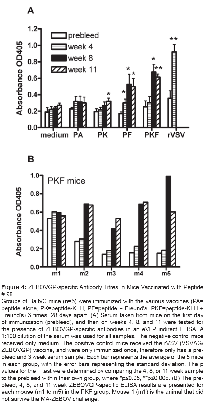
As part of the protection study the humoural and T cell mediated immune responses were examined in the mice, leading up to the MA-ZEBOV challenge. An eVLP indirect ELISA was performed to determine the level of the ZEBOVGP-specific antibodies in the serum of the mice at day 0 (prebleed), and at 4, 8, and 11 weeks post-immunization (Figure 4A). As with the negative control mice that only received the medium, the PA group did not produce any ZEBOVGP-specific antibodies. When the peptide was bound to KLH (PK mice), a significant level of antibodies was seen by week 11. The addition of Freund’s adjuvant to either the peptide alone (PF) or the peptide-KLH conjugation (PKF) resulted in high antibody levels after only two immunizations. The positive control mice that received one injection of rVSV produced very high antibody titres. The levels of ZEBOVGP-specific antibodies in the PF and PKF groups were 57% and 74% respectively, after 2 immunizations, and 54% and 67% after 3 immunizations, of the titres seen with the rVSV positive control group. However, despite the higher titres in the PKF group one mouse did not survive whereas all mice survived in the PF group. When examining the titres in each mouse of the PKF group (Figure 4B), the mouse that did not survive (m1) had high background titres for the prebleed and the titres did not rise above background after immunization, suggesting an ineffective ZEBOVGP-specific humoural response to the immunization. Overall, immunization with peptide #98 did result in the production of ZEBOVGP-specific antibodies, but the strength of the antibody response was dependent upon the carrier molecules and adjuvant used.
Figure 4: ZEBOVGP-specific Antibody Titres in Mice Vaccinated with Peptide
# 98.
Groups of Balb/C mice (n=5) were immunized with the various vaccines (PA=
peptide alone, PK=peptide-KLH, PF=peptide + Freund’s, PKF=peptide-KLH +
Freund’s) 3 times, 28 days apart. (A) Serum taken from mice on the first day
of immunization (prebleed), and then on weeks 4, 8, and 11 were tested for
the presence of ZEBOVGP-specific antibodies in an eVLP indirect ELISA. A
1:100 dilution of the serum was used for all samples. The negative control mice
received only medium. The positive control mice received the rVSV (VSVΔG/
ZEBOVGP) vaccine, and were only immunized once, therefore only has a prebleed
and 3 week serum sample. Each bar represents the average of the 5 mice
in each group, with the error bars representing the standard deviation. The p
values for the T test were determined by comparing the 4, 8, or 11 week sample
to the prebleed within their own group, where *p≤0.05, **p≤0.005. (B) The prebleed,
4, 8, and 11 week ZEBOVGP-specific ELISA results are presented for
each mouse (m1 to m5) in the PKF group. Mouse 1 (m1) is the animal that did
not survive the MA-ZEBOV challenge.
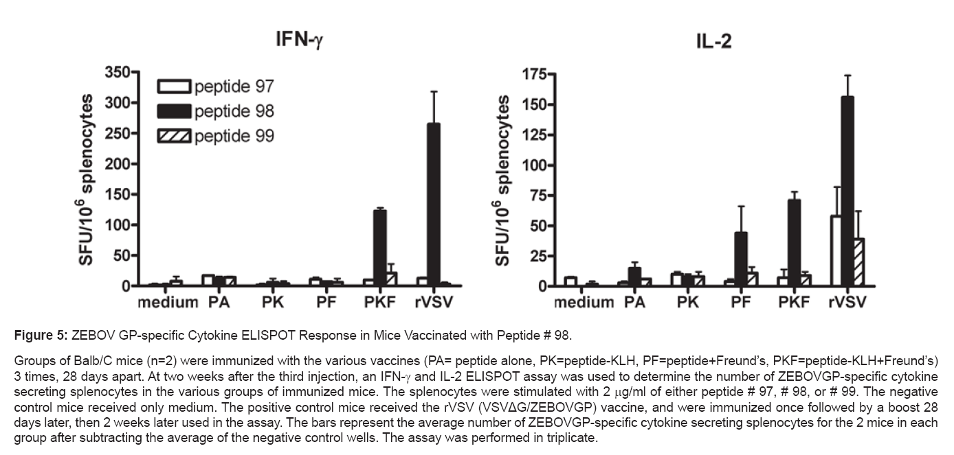
In addition to the humoural response the ZEBOVGP-specific T cell mediated response was determined using an IFN-g and IL-2 ELISPOT assay (Figure 5). Two weeks after the third immunization, the spleens of 2 mice from each group were tested in order to determine the number of splenocytes responding to peptide # 98, or to two neighbouring peptides, # 97 and # 99. For all three peptides, there were no IFN-g or IL-2 secreting splenocytes in the medium, PA, or PK groups. The PF mice did not respond to any of the three peptides in the IFN-g assay, but did respond to peptide #98 in the IL-2 assay. In comparison the PKF group responded only to peptide #98 in both the IFN-g and IL-2 assay. The positive control rVSV immunized mice had a large number of splenocytes secreting IFN-g to peptide #98, and secreted IL-2 predominantly to peptide #98 with a much weaker response to peptides # 97 and # 99. Overall, the strongest T cell responses were seen in the groups receiving Freund’s adjuvant and they were highly specific for peptide # 98.
Figure 5: ZEBOV GP-specific Cytokine ELISPOT Response in Mice Vaccinated with Peptide # 98.
Groups of Balb/C mice (n=2) were immunized with the various vaccines (PA= peptide alone, PK=peptide-KLH, PF=peptide+Freund’s, PKF=peptide-KLH+Freund’s)
3 times, 28 days apart. At two weeks after the third injection, an IFN-g and IL-2 ELISPOT assay was used to determine the number of ZEBOVGP-specific cytokine
secreting splenocytes in the various groups of immunized mice. The splenocytes were stimulated with 2 mg/ml of either peptide # 97, # 98, or # 99. The negative
control mice received only medium. The positive control mice received the rVSV (VSVΔG/ZEBOVGP) vaccine, and were immunized once followed by a boost 28
days later, then 2 weeks later used in the assay. The bars represent the average number of ZEBOVGP-specific cytokine secreting splenocytes for the 2 mice in each
group after subtracting the average of the negative control wells. The assay was performed in triplicate.
Discussion
A cytokine ELISPOT assay was used to determine the immunodominant epitope of the ZEBOV glycoprotein. As a reliable assay for determining memory cell immune responses in vaccine clinical trials, the ELISPOT assay is a sensitive and reproducible test for determining the antigen-specific T cell response at the single cell level. Determination of the frequency of the T cell response reflects the in vivo cellular frequency [15,16]. Additionally, the ELISPOT assay can be used to determine what cytokine profile is induced. In this study, Balb/C mice were immunized with the VSVΔG/ZEBOVGP vaccine in which the VSV glycoprotein was replaced by the ZEBOV GP. Peptides spanning the ZEBOV GP were used to stimulate the splenocytes from these mice in 5 different cytokine ELISPOT assays in order to determine the ZEBOVGP-specific T cell cytokine profile. Upon exposure to the peptides the strongest responses were demonstrated in the Th1 cytokines IFN-g, TNF, and IL-2, whereas the two Th2 cytokine responses (IL- 4 and IL-10) were negligible. This suggests that ZEBOVGP presented in the context of VSVΔG/ZEBOVGP during immunization produced ZEBOVGP-specific memory T cells secreting Th1 cytokines. This is an expected and typical immune response to viral infections. Of the three Th1 cytokines, the frequency of IFN-g producing splenocytes was 2.7 times higher than for IL-2 and TNF. Having a strong IFN-g response is vital for control of ZEBOV infections as ZEBOV has been shown to shut down IFN-g [17-19]. Therefore restoring a rapid IFN-g response early during an infection would be vital in countering the effects of a ZEBOV infection and tipping the scale towards survival.
IFN-g has strong antiviral effects that can inhibit viral replication directly, as well as stimulate host cellular immune response genes [20]. IFN-g is produced by memory and effector T cells but not naïve cells making the IFN-g ELISPOT assay a valuable tool for examining effector T cells responses in immunized animals [21]. In the VSVΔG/ ZEBOVGP immunized mice a higher percentage of ZEBOVGPspecific splenocytes were secreting IFN-g therefore the IFN-g ELISPOT assay was used to determine the immunodominant ZEBOVGP epitope. The frequency of ZEBOVGP-specific splenocytes did not span the entire region of GP. The initial cytokine profile assays demonstrated that ZEBOVGP peptide pool # 2 induced overwhelmingly the highest number of spot forming units (SFU) for all three Th1 cytokine secreting splenocytes. To further define the immunodominant epitope, the peptides from pool # 2 were split into another set of pools according to a matrix in order to determine which specific peptides were inducing the IFN-g T cell response. Of the four possible peptides identified (# 72, 74, 96, and 98) only peptide # 98 stimulated a strong IFN-g response suggesting peptide #98 was the immunodominant epitope. This peptide HNTPVYKLDISEATQ (amino acids 389-403), which is conserved across both the Mayinga and Kikwit strains, is located in the mucin domain of GP. The mucin domain is believed to be responsible for the cytopathicity in GP expressing cells lines, and may play a critical role in the pathogenesis of filoviral disease [22-24]. Interestingly, protective monoclonal antibody 6D8-1-2 also binds this epitope which suggests that this epitope may elicit both a humoural and a cellular immune response [25].
Finding a strongly immunogenic epitope in a protein does not guarantee that using the peptide in a vaccine can prevent disease. Therefore, in order to demonstrate the capacity of peptide # 98 to induce protective immunity, mice were immunized with the peptide alone (PA), peptide conjugated to KLH (PK), or peptide along with Freund’s adjuvant (PF and PKF). Small peptides and proteins are not very effective at eliciting a strong immune response therefore it is not unexpected that peptide alone did not increase survival from a lethal MA-ZEBOV infection. Therefore, KLH was employed as a carrier for the peptide in order to make the peptide more immunogenic [26]. While the KLH-conjugated peptide (PK) increased the mean time to death it did not prevent death. However, this model used a high dose challenge and it is possible that there could have been an improved rate of survival with a lower challenge dose of MA-ZEBOV. A more potent mechanism for enhancing the immune response is to inject the peptide in the presence of an adjuvant such as Freund’s [27]. The adjuvanted peptide was extremely effective as it increased survival to 100%. This demonstrates that peptide #98 induces a protective immune response.
The humoural and T cell mediated immune response elicited by peptide #98 was compared for each of the various vaccine protocols. The medium negative control, and the peptide alone (PA) immunization did not stimulate any immune response. This is not surprising as small peptides are very weak immunogens. One possible reason is that the peptide vaccine contains just one epitope and therefore may induce only a very narrow and weak response. Or, peptides in aqueous solutions may induce tolerance as the peptide does not contain “danger signals” that are required to induce inflammatory responses needed to stimulate the immune system [28]. Therefore, an adjuvant or carrier is often required to induce this response (reviewed in [29]. Adding the carrier protein KLH to the peptide (PK) resulted in a weak ZEBOVGPspecific antibody response at week 11 but no noticeable T cell response. This is reflected in the PK vaccines ability to increase the mean time to death but not improve survival. The addition of Freund’s adjuvant to the peptide (PF and PKF) provided the necessary signals which resulted in a strong ZEBOVGP-specific antibody response after just two immunizations; and splenocytes secreting IL-2 (PKF and PF) or IFN-g (PKF) after 3 immunizations. The T and B cell response is not as strong as what is observed in mice immunized with the VSVΔG/ ZEBOVGP vaccine, most likely due to several reasons. Firstly, the antigenic dose of the VSVΔG/ZEBOVGP vaccine would be higher as it is a live replicating virus. Secondly, as VSVΔG/ZEBOVGP includes the entire ZEBOV GP, it contains more B and T cell epitopes than the peptide vaccine, and would thus provide a broader antigenic stimulus. This is reflected in the IL-2 ELISPOT results where there were IL-2 secreting splenocytes specific for peptide # 98 as well as peptides # 97 and # 99. Finally, the adjuvants “danger signals” provided by VSVΔG/ ZEBOVGP will be quantitatively and qualitatively different than those provided by Freund’s. Surprisingly, the PKF group which had a stronger B- and T- cell response than the PK group, had one mouse (m1) die. This mouse was later found to have a weak/absent ZEBOVGP-specific antibody response, as the antibody titres never exceeded the initially high background levels. There are no corresponding ELISPOT results for this mouse as the ELISPOT results come from mice not included in the challenge. However, previous work has shown that CD8+ T cells are not required for survival yet passive transfer of ZEBOVGP-specific antibodies does provide protection [14]. Therefore, it is possible that the one mouse that died did not have a sufficient antibody response against the ZEBOV GP, for reasons unknown. The PF group which did not have any IFN-g secreting splenocytes did have complete survival. This evidence supports the necessity for antibodies in promoting survival during a ZEBOV infection.
The peptide sequence HNTPVYKLDISEATQ is conserved across the Mayinga and Kikwit strains of Zaire Ebolavirus, but is not conserved across the various species of Ebola. Therefore this peptide may not provide cross-protection against other Ebola virus species. The peptide HNTPVYKLDISEATQ is not a previously identified CD8+ CTL epitope. Two other H-2d restricted peptides that have been identified in Balb/C mice include VSTGTGPGAGDFAFHK (amino acids 141-155) identified using the Venezuelan Equine Encephalitis Virus Replicon (VRP) expressing the Zaire glycoprotein as a vaccine [30], and the LYDRLASTI (amino acids 161-169) identified using the liposome plus irradiated Ebola virus vaccine (L(EV)) [31]. Both of these studies immunized with vaccines containing the complete glycoprotein and then either identified the immunodominant CTL epitope using overlapping peptides, or were tested against peptides predicted by software to be strong binders for H-2d, respectively. Possible reasons why each study did not pick up the others immunodominant peptides may be due to the fact that all three approaches used different assays to identify the peptides, as well as different vaccine delivery systems. In the Olinger VRP-GP vaccine study, the VSTGTGPGAGDFAFHK epitope was identified using intracellular cytokine staining (ICS) by flow cytometry, yet the epitope was undetected in their IFN-g ELISPOT assay. This is consistent with our results where we also did not detect their epitope in our IFN-g ELISPOT assay, and we did not perform an ICS assay. The L(EV) vaccine used a software to determine the binding predictions for H-2d based on the amino acid sequence of ZEBOV GP. Because their predicted peptide sequence did not match that of our peptide # 98, our peptide was not part of their ELISPOT, or Cr51 CTL assay. We however, did not identify their predicted epitope in our ELISPOT. This may be because using a peptide matrix pool where there are 8 peptides in one pool, may result in the response to one peptide being overwhelmed by the response to another peptide. Comparison of the various assays to define immunodominant epitopes needs to be addressed to determine which method might predict the most effective epitope.
It is difficult to say which epitope will be most effective in providing protection in an in vivo model. However, the current study is the first to demonstrate that the 15 amino acid peptide based on the identified epitope can provide protection in a mouse model. The other two studies identified their epitopes as either CD4+ or CD8+ specific. In our study the ELISPOT assay was not able to differentiate between CD4+ or CD8+ T lymphocytes, therefore we used a web based epitope prediction software (IEBC Analysis Resource at https://tools.immuneepitope.org/main/) and identified epitopes within HNTPVYKLDISEATQ that bound to MHCI and MHCII with an IC50 of approximately 30 mM, and 4-7 mM, respectively. This suggests that the peptide has a 4-8 times higher binding affinity for MHCII than MHCI. Activation of CD4+ T lymphocytes would suggest the activation of a humoural immune response. Interestingly, our peptide was also found to be bound by monoclonal antibody 6D8-1-2 [25].The identification of distinctly different epitopes using 3 different vaccine platforms may be due to differences in the route of immunization as well as the vaccine formulation. The L(EV) vaccine generated a CTL response, but the irradiated EV in absence of the liposome did not, suggesting that the additional signals, or route of cellular entry provided by the liposome can influence the immune response. Liposome encapsulated antigen is presumably processed by a different intracellular pathway which could possibly lead to a different immune response [32]. The danger signals presented by the other components of each vaccine would also be distinctly different which may manifest in different cytokines being secreted or different cells being activated. Additional research into the protective mechanisms has to be determined for vaccines and Ebola virus infections.
All of the data taken together suggests that peptide # 98 can induce both a T and B cell immune response that is sufficient to protect against a high dose lethal challenge of MA-ZEBOV. The level of induction of the B and T cells is not as strong as what is observed with the rVSV vaccine. However, the peptide contains only one epitope whereas the rVSV vaccine contains numerous epitopes. The fact that a single peptide can provide sufficient protection suggests that with a suitable delivery system and adjuvant, a subunit vaccine containing the single peptide could be an effective vaccine that is safer than the live replicating virus vaccine. Although the antibody titres were high there is evidence that Th1 cell mediated immune response may also play an important role in providing protection against a ZEBOV infection. The cytokine profile suggested a Th1 cytokine response, and since the IFN-g ELISPOT was used to determine the immunodominant epitope it is plausible that the cell mediated immune response is also a factor. The ELISPOT assay used here can only quantitate an overall T cell response, and does not determine whether the ZEBOVGP-specific response was mediated by CD4+ or CD8+ T cells. Future experiments should include a flow cytometry based intracellular cytokine assay that can differentiate which T lymphocyte recognizes the immunodominant epitope # 98 identified here. Additionally, further investigation in to the relative contribution of the T and B cell response towards protection by peptide # 98 could be determined through the adoptive T cell or antibody transfer studies, or through the vaccination of T- or B-cell deficient mice. Identifying the cytokine profile and immunodominant ZEBOVGP epitope in the VSVΔG/ZEBOVGP vaccine aids in determining the correlates of immune protection against ZEBOV, and will play an important role in developing assays to establish the efficacy of the vaccine in eliciting a protective immune response in vaccinated individuals. The VSVΔG/ ZEBOVGP vaccine completely protects non-human primates against a lethal high dose challenge indicating that GP is sufficient to elicit protection [1-3]. While using VSV as the vector carrying ZEBOVGP to immunize against a ZEBOV infection, the identification of the cytokine profile and immunodominant epitope will also allow for the development of future subunit vaccines using peptide #98.
Acknowledgements
We would like to thank the Veterinary Technical Services at the National Microbiology Laboratory in Winnipeg for their animal work, and Jonathan Audet for his help with the manuscript. The source of funding for this project is from the Chemical, Biological, Radiological-nuclear, and Explosives Research and Technology Inititative (CRTI), and the Public Health Agency of Canada (PHAC).
References
- Garbutt M, Liebscher R, Wahl-Jensen V, Jones S, Moller P, et al. (2004) Properties of replication-competent vesicular stomatitis virus vectors expressing glycoproteins of filoviruses and arenaviruses. J Virol 78: 5458-5465.
- Jones SM, Feldmann H, Stroher U, Geisbert JB, Fernando L, et al. (2005) Live attenuated recombinant vaccine protects nonhuman primates against Ebola and Marburg viruses. Nat Med 11: 786-790.
- Qiu X, Fernando L, Alimonti JB, Melito PL, Feldmann F, et al. (2009) Mucosal immunization of cynomolgus macaques with the VSVDeltaG/ZEBOVGP vaccine stimulates strong ebola GP-specific immune responses. PLoS One 4: e5547.
- Geisbert TW, Jahrling PB (2003) Towards a vaccine against Ebola virus. Expert Rev Vaccines 2: 777-789.
- Warfield KL, Swenson DL, Olinger GG, Kalina WV, Aman MJ, et al. (2007) Ebola virus-like particle-based vaccine protects nonhuman primates against lethal Ebola virus challenge. J Infect Dis 196 Suppl 2: S430-7.
- Pass HA, Schwarz SL, Wunderlich JR, Rosenberg SA (1998) Immunization of patients with melanoma peptide vaccines: immunologic assessment using the ELISPOT assay. Cancer J Sci Am 4: 316-323.
- Vasan S, Schlesinger SJ, Huang Y, Hurley A, Lombardo A, et al. (2010) Phase 1 safety and immunogenicity evaluation of ADVAX, a multigenic, DNA-based clade C/B' HIV-1 candidate vaccine. PLoS One 5: e8617.
- Lewis JJ, Janetzki S, Schaed S, Panageas KS, Wang S, et al. (2000) Evaluation of CD8(+) T-cell frequencies by the Elispot assay in healthy individuals and in patients with metastatic melanoma immunized with tyrosinase peptide. Int J Cancer 87: 391-398.
- Gebauer BS, Hricik DE, Atallah A, Bryan K, Riley J, et al. (2002) Evolution of the enzyme-linked immunosorbent spot assay for post-transplant alloreactivity as a potentially useful immune monitoring tool. Am J Transplant 2: 857-866.
- Schnell MJ, Buonocore L, Kretzschmar E, Johnson E, Rose JK (1996) Foreign glycoproteins expressed from recombinant vesicular stomatitis viruses are incorporated efficiently into virus particles. Proc Natl Acad Sci USA 93: 11359- 11365.
- Bray M, Davis K, Geisbert T, Schmaljohn C, Huggins J (1998) A mouse model for evaluation of prophylaxis and therapy of Ebola hemorrhagic fever. J Infect Dis 178: 651-661.
- Wahl-Jensen V, Kurz SK, Hazelton PR, Schnittler HJ, Stroher U, et al. (2005) Role of Ebola virus secreted glycoproteins and virus-like particles in activation of human macrophages. J Virol 79: 2413-2419.
- Wahl-Jensen VM, Afanasieva TA, Seebach J, Stroher U, Feldmann H, et al. (2005) Effects of Ebola virus glycoproteins on endothelial cell activation and barrier function. J Virol 79: 10442-10450.
- Jones SM, Stroher U, Fernando L, Qiu X, Alimonti J, et al. (2007) Assessment of a vesicular stomatitis virus-based vaccine by use of the mouse model of Ebola virus hemorrhagic fever. J Infect Dis 196 Suppl 2: S404-12.
- Currier JR, Kuta EG, Turk E, Earhart LB, Loomis-Price L, et al. (2002) A panel of MHC class I restricted viral peptides for use as a quality control for vaccine trial ELISPOT assays. J Immunol Methods 260: 157-172.
- Kreher CR, Dittrich MT, Guerkov R, Boehm BO, Tary-Lehmann M (2003) CD4+ and CD8+ cells in cryopreserved human PBMC maintain full functionality in cytokine ELISPOT assays. J Immunol Methods 278: 79-93.
- Bray M , Mahanty S (2003) Ebola hemorrhagic fever and septic shock. J Infect Dis 188: 1613-1617.
- Bray M and Geisbert TW (2005) Ebola virus: the role of macrophages and dendritic cells in the pathogenesis of Ebola hemorrhagic fever. Int J Biochem Cell Biol 37: 1560-1566.
- Harcourt BH, Sanchez A, Offermann MK (1999) Ebola virus selectively inhibits responses to interferons, but not to interleukin-1beta, in endothelial cells. J Virol 73: 3491-3496.
- Schroder K, Hertzog PJ, Ravasi T, Hume DA (2004) Interferon-gamma: an overview of signals, mechanisms and functions. J Leukoc Biol 75: 163-189.
- Zimmermann C, Prevost-Blondel A, Blaser C, Pircher H (1999) Kinetics of the response of naive and memory CD8 T cells to antigen: similarities and differences. Eur J Immunol 29: 284-290.
- Yang ZY, Duckers HJ, Sullivan NJ, Sanchez A, Nabel EG, et al. (2000) Identification of the Ebola virus glycoprotein as the main viral determinant of vascular cell cytotoxicity and injury. Nat Med 6: 886-889.
- Takada A, Watanabe S, Ito H, Okazaki K, Kida H, et al. (2000) Downregulation of beta1 integrins by Ebola virus glycoprotein: implication for virus entry. Virology 278: 20-26.
- Simmons G, Wool-Lewis RJ, Baribaud F, Netter RC, Bates P (2002) Ebola virus glycoproteins induce global surface protein down-modulation and loss of cell adherence. J Virol 76: 2518-2528.
- Wilson JA, Hevey M, Bakken R, Guest S, Bray M, et al. (2000) Epitopes involved in antibody-mediated protection from Ebola virus. Science 287: 1664- 1666.
- Harris JR, Markl J (1999) Keyhole limpet hemocyanin (KLH): a biomedical review. Micron 30: 597-623.
- Heegaard PM, Dedieu L, Johnson N, Le Potier MF, Mockey M, et al. (2010) Adjuvants and delivery systems in veterinary vaccinology: current state and future developments. Arch Virol 156: 183-202.
- Kyburz D, Aichele P, Speiser DE, Hengartner H, Zinkernagel RM, et al. (1993) T cell immunity after a viral infection versus T cell tolerance induced by soluble viral peptides. Eur J Immunol 23: 1956-1962.
- Storni T, Kundig TM, Senti G, Johansen P (2005) Immunity in response to particulate antigen-delivery systems. Adv Drug Deliv Rev 57: 333-355.
- Olinger GG, Bailey MA, Dye JM, Bakken R, Kuehne A, et al. (2005) Protective cytotoxic T-cell responses induced by venezuelan equine encephalitis virus replicons expressing Ebola virus proteins. J Virol 79: 14189-14196.
- Rao M, Bray M, Alving CR, Jahrling P, Matyas GR (2002) Induction of immune responses in mice and monkeys to Ebola virus after immunization with liposome-encapsulated irradiated Ebola virus: protection in mice requires CD4(+) T cells. J Virol 76: 9176-9185.
- Peachman KK, Rao M, Alving CR, Palmer DR, Sun W, et al. (2005) Human dendritic cells and macrophages exhibit different intracellular processing pathways for soluble and liposome-encapsulated antigens. Immunobiology 210: 321-333.
Relevant Topics
- Anthrax Bioterrorism
- Bio surveilliance
- Biodefense
- Biohazards
- Biological Preparedness
- Biological Warfare
- Biological weapons
- Biorisk
- Bioterrorism
- Bioterrorism Agents
- Biothreat Agents
- Disease surveillance
- Emerging infectious disease
- Epidemiology of Breast Cancer
- Information Security
- Mass Prophylaxis
- Nuclear Terrorism
- Probabilistic risk assessment
- United States biological defense program
- Vaccines
Recommended Journals
Article Tools
Article Usage
- Total views: 15344
- [From(publication date):
specialissue-2011 - Nov 17, 2025] - Breakdown by view type
- HTML page views : 10554
- PDF downloads : 4790

