Short Communication Open Access
Anterior Approach for Ultrasound-guided Pudendal Block
| Teresa Parras*, Rafael Blanco and Beenu Madhavan | |
| St George's Hospital, London, UK | |
| *Corresponding Author : | Teresa Parras St George's Hospital, London, UK Tel: 00447776433710 E-mail: tparrasmaldonado@gmail.com |
| Received: September 10, 2015; Accepted: February 04, 2016; Published: February 10, 2016 | |
| Citation: Parras T, Blanco R, Madhavan B (2016) Anterior Approach for Ultrasound-Guided Pudendal Block. J Pain Relief 5:230. doi:10.4172/2167-0846.1000230 | |
| Copyright: © 2016 Parras T, et al. This is an open-access article distributed under the terms of the Creative Commons Attribution License, which permits unrestricted use, distribution, and reproduction in any medium, provided the original author and source are credited. | |
Visit for more related articles at Journal of Pain & Relief
Abstract
We present a descriptive study, to perform a noninvasive ultrasound-guided scanning technique that allows repeatable visualization of the pudendal nerve, in its anterior approach. We scanned 56 perineal areas of 28 volunteers bilaterally, analyzing the localization of the pudendal nerve and anatomy of structures around it. Our results shows that the easier way to find the nerve is placing the probe transversal to the line between scrotum or mons pubis and the anal margin, medial to ischial tuberosity; identifying the nerve running along the transverse perineal muscle and medial to pudendal artery. The ultrasound-guided anterior approach of pudendal nerve block is an easy and reliable performing technique, avoiding neuroestimulation or other invasive techniques.
| Keywords |
| Nerve block; Analgesia; Pudendal; Pain |
| Introduction |
| A pudendal nerve block is a safe alternative to caudal anaesthesia to provide anaesthesia and/or analgesia for the perineum. It can be considered for a variety of clinical indications in general surgery, obstetrics, urology and chronic pain. The pudendal nerve block has evolved since its first description with respect to techniques (fluoroscopy, neurostimulation and ultrasound) and anatomic references. Historically the pudendal nerve block was performed by surgeons and obstetricians, but has gradually gone out of favor, as the block was not always reliable. |
| The pelvic floor consists of muscles, ligaments and fascia to form a hammock that supports the abdomino-pelvic viscera at the base of the pelvis. It is composed of layers of muscles that connect the anterior and posterior pelvic ring and can be separated into the superficial and deep floors. The superficial pelvic floor includes the bulbospongiosus, ischiocavernosus, and superficial transverse perineus muscles. The deep transverse perineous muscle is present in the deep pelvic floor, which has two major components, the pubovisceral, (composed of the pubococcygeus and puborectalis muscles), and the iliococcygeal muscles. |
| The major muscle in the pelvic floor is the levator ani muscle and its integrity plays a key role in maintenance of pelvic organ support. In women, this muscle complex surrounds the urogenital hiatus that contains the urethra, vagina and anal canal. |
| In the perineal area there are two triangles, the anterior or urogenital triangle and posterior or anal triangle (Figure 1). The lateral and medial borders of anterior triangle are composed of the ischiocavernosus and bulbospongiosus muscle, and the base by the transverse perineal muscle. The base of the posterior triangle is again the transverse perineal muscle, with the sphincter ani and sacrotuberosus ligament as the medial and lateral borders respectively [1-3]. |
| A transverse line coursing from the ischial tuberosities through the perineal body sets the stage for separating the two triangles. |
| Urogenital or anterior triangle: this extends from the ischial tuberosities to the pubic symphysis. It contains the root of the scrotum and penis in males and the vulva of females. |
| Anal or posterior triangle: this extends from the ischial tuberosities to the coccyx. It contains the anal canal and its orifice, the anus. |
| The perineal body or the central tendon of the perineum, lies between the vagina and the anus and comprises of fibrous tissue and the superficial pelvic floor musculature. |
| For many years, authors have stated that the levator ani had dual innervation from the pudendal nerve and also direct branches from the S3 and S4 motor roots. The posterior part of the pelvic floor is innervated directly by efferents from the S2-4 nerve roots, whereas the pudendal nerve and its three branches (the dorsal nerve of the penis or clitoris, the perineal branch, and the inferior hemorrhoidal nerve) innervate the anterior pelvic floor [4]. |
| In order to understand the ultrasound technique of blocking the pudendal nerve it is paramount to know its course in the pelvis and its neighbouring anatomy. The pudendal nerve is formed from the ventral primary rami of S2, S3, and S4. It then forms the neurovascular pudendal bundle with the internal pudendal artery and venous plexus [5]. |
| As it enters the pudendal canal or Alcock’s canal, the pudendal nerve gives the inferior rectal nerve providing motor supply to the external anal sphincter, sensation to the skin anterior and lateral to the anus, and also the mucosa of the lower part of the anal canal. In the ischiorectal fossa, the pudendal nerve gives two branches, the perineal nerve, and the dorsal nerve of the penis or clitoris. The perineal nerve divides into the superficial and deep perineal nerves. The superficial branch (called as posterior scrotal or labial branches) provides the sensation to the skin of the posterior aspect of scrotum or labia. The deep branch gives motor innervation to the bulbospongiosus, ischiocavernosus, transverse perineal muscles of urogenital triangle. The penile or clitorial skin is innervated by the dorsal nerve of the penis or clitoris, which travels ventrally under the symphysis pubis. The dorsal surface at the base of penis or the mons pubis and labia majora is supplied by the ilioinguinal nerve. |
| Literature provides several techniques to block the pudendal nerve, all of which depend on localizing the anatomic landmarks around the nerve [6-8]. The most common approach has been fluoroscopic guidance, where the needle is aimed to be placed as close as possible to the ischial spine. The flaw in this method is that the pudendal nerve is in the interligamentous plane, which cannot be visualized by fluoroscopy. An alternative method using CT (computerized tomography) to identify the interligamentous plane has been described before [5]. There are a number of limitations including the fact that it is performed without real-time visual control and also is exclusively avalaible to radiologists, not to mention the exposure of radiation. |
| Ultrasound imaging is becoming popular as the imaging method in the investigation of pelvic floor disorders, current approaches include transabdominal, transperineal/translabial, transvaginal/introital or transrectal. Real-time transperineal sonography has enhanced the appreciation of morphology and dynamics of the pelvic floor. Standard images are obtained from longitudinal and axial planes by placing the transducer between the vagina or scrotum and anus. |
| Ultrasound guided nerve block have advantages including detecting the spread of the injected solution close to real-time, visualization of the pudendal artery, and avoidance of ionizing radiation. Ultrasonography can improve the accuracy and safety and therefore minimize the chance of failure and complications. It is the most widely available imaging modality outside of radiology. Introduction of novel transducers and sophisticated software to the market has resulted in units that provide superior image than before of the anatomy of pelvic floor. Progress of this technology allows visualization of the bony, vascular and soft tissue structures that are essential in the success of the block, these include the ischial spine, the internal pudendal artery and perineal transverse muscle. |
| Case Presentation |
| We present a descriptive study to perform a noninvasive ultrasoundguided scanning technique, that allows easy repeatable visualization of the pudendal nerve. We describe the anterior approach, where effective block depends on accurate localization of the pudendal nerve, being essential to have knowledge and understand the sonoanatomy of the nerve and the structures around it. |
| Methods |
| We present a descriptive study of 28 volunteers, with the written informed consent, to perform the ultrasound-guided perineal scanning, observing 56 pudendal areas. |
| Using a linear probe (8-15 MHz) or a hockey stick (6-13 MHz) we can visualize the bulbospongiosus, ischiocavernosus and transverse perineal muscles, pubis, ischial spine, and the pudendal artery. |
| The pubovisceral muscle lies hidden in the pelvis and until recently magnetic resonance imaging was the only imaging method capable of assessing it in the axial plane. The lateral aspect of the puborectalis muscle can be difficult to define, due to the difficulty in distinguishing between puborectalis and pubococcygeus. Technological advances in three- and four-dimensional perineal ultrasound have made available a new diagnostic tool, which, with multiplanar imaging, makes it possible to visualize the pubovisceral muscle in the axial plane. Using a 5 MHz ultrasound probe placed at the vaginal introitus and orientated posteriorly, the puborectalis can be recognized in the sagittal plane as a hyperechogenic structure posterior to the proximal third of the anal canal. |
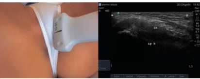
| The transperineal approach is performed with the patient in lithotomy position, with the help of a cushion underneath the pelvis was lifted and rotated ventrally, hips flexed and knees bent with the legs abducted and slightly rotated outwards. Visualization is best achieved by placing the ultrasound probe at the perineum between the scrotum or clitoris and the anus (Figure 2 left). The transducer can usually be placed firmly on the perineum and the symphysis pubis without causing discomfort, unless there is marked atrophy or local skin changes. The image will also depend on the hydration state of tissues, which generally is best in pregnancy and poorest in elderly women with marked atrophy. The resulting image shows the symphysis pubis anteriorly to the anorectal junction posteriorly. |
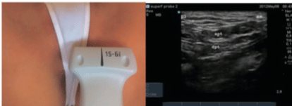
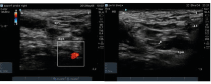
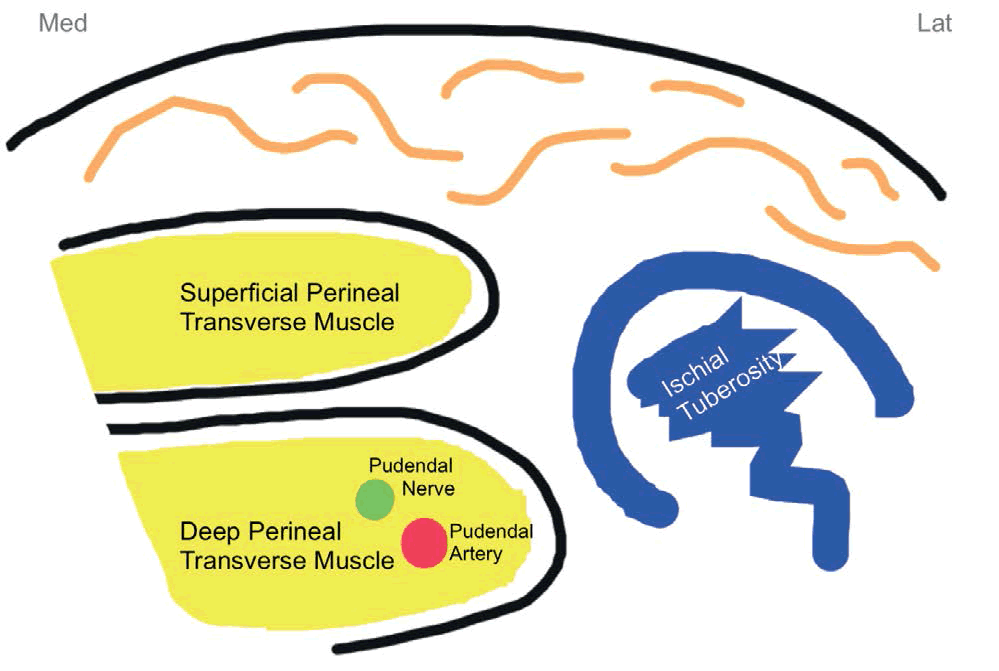
| The recommended technique is to start by identifying the reference structures. At first, place the probe in the longitudinal axis at the anterior triangle, with the symphysis pubis anteriorly and the perineal body posteriorly (Figure 2 left). At this level identify the ischiopubic ramus and the ischiocavernosus muscle lying above (Figure 2 right). Next, place the probe in the transverse axis of the perineal body to identify the superficial and deep transverse perineal muscles (Figure 3). Then move the probe laterally to visualize the posterior edge of the ischiopubic ramus. Medial to that, we find the internal pudendal artery, which can be confirmed with colour Doppler (Figure 4 left). A longitudinal or transverse view of the artery can be obtained depending on the probe position. Placing the probe in a transverse position, identifying the ischial tuberorisity, we visualize the pudendal artery and perineal transverse muscles, and the perineal nerve, medial to the artery (Figure 4 right). |
| Results |
| We scanned 23 females and 5 males bilaterally, with an average of 37.78 years, 74.3 kg. |
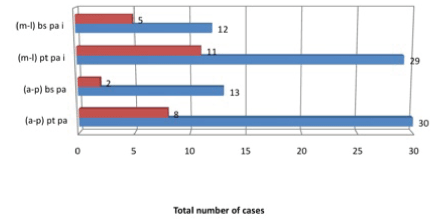
| We analyze the position of the pudendal artery in relation with muscles (bulbospongiosus and perineal transverse) in transverse and longitudinal probe position. Placing the probe in transverse position, we identify pudendal artery between bulbospongious and ischium in 12 images, and we visualize the nerve in 5 images, while we find the artery between perineal transverse muscle and ischium in 29 cases, and the nerve in 11. And if we place the probe in longitudinal, we identify the artery posterior to bulbospongiosus muscle in 13 images and we just find the nerve in 2, though the artery is positioned posterior to perineal transverse in 30 images and the nerve can be seen in 8. |
| Therefore, placing the transducer in transverse position we identified the nerve in a total of 16 patients but in longitudinal position we identified it in 8 patients. With this results we conclude that if we place our probe in transverse axis (transversal to the line between the scroutm or mons pubis and the anal margin, medial to ischial tuberosity) it will be easier to identify pudendal artery; and medial to it, the pudendal nerve, along the perineal transverse muscle (Figure 5). |
| The sonoanatomy with the transducer in this position is show in Figure 6. |
| Discussion |
| The difficulty in visualizing the pudendal nerve by ultrasound had been previously reported in literature [4]. At the level of the ischial spine the pudendal nerve is 4 to 6 mm in diameter. Also, at this level in 30-40% of cases, the pudendal nerve would have already divided into 2 or 3 branches. The difficulty in isolating the pudendal nerve is due the surrounding connective and fatty tissue. The injection of local anaesthesic can improve the visualization of the nerve under ultrasound [9]. |
| The pudendal nerve block is used for anaesthesia or analgesia for perineal surgery, hemorrhoidectomy, perineal neuralgia, neurogenic bladder dysfunction and chronic pelvic pain causes by pudendal nerve entrapment. In males it can be used for transrectal prostate biopsy, circumcision, penile prothesis operations, and in females for colpoperineorrhaphy [10]. |
| Most recent studies on pudendal nerve block have been conducted mainly in urological or proctological procedures [11]. Its use in obstetrics however to date has been poorly studied. In this field, it is used for pain relief during the second stage of labor in spontaneous vaginal or instrumental deliveries (vacuum or forceps), for episiotomy and repair of severe lacerations (recto-vaginal fistulae, suture of perineum). Multipara women with pain at delivery end, can especially benefit from the use of a pudendal nerve block. It is another method that an obstetrician could offer a parturient, a second line choice after neuraxial block with general anaesthesia as the last alternative in acute emergencies and in cases of failed regional anaesthesia or when the mother refuses any other anaesthesia despite proper information or proves unable to cooperate. |
| Episiotomy or tearing of perineal tissues during childbirth is associated with significant pain in the postpartum period. Although the use of episiotomy is often debated, it remains the most common surgical procedure experienced by women [10]. Even though pain from episiotomy may be severe and can result in significant discomfort it is poorly treated [12]. |
| The American College of Obstetricians and Gynecologists defines chronic pelvic pain as “non-cyclic pain of 6 or more months duration that localizes to the anatomic pelvis, anterior abdominal wall at or below the umbilicus, the lumbosacral back, or the buttocks and is of sufficient severity to cause functional disability or lead to medical care”. Chronic pelvic pain is a clinical diagnosis made in patients with the typical history involving the penis or clitoris, scrotum or labia, perineum and anorectal region. This may arise from pudendal nerve entrapment. To exclude this pathology is crucial to confidently block the pudendal nerve to guide for the management. |
| Conclusion |
| Pelvic floor sonography is a relatively simple, noninvasive, and reliable method to assess not only functionality but also morphology, this pudendal anterior approach described could be used to perform analgesic or anesthetic blocks. The potential risks for pudendal block include hematoma, infection, nerve damage as well as local anesthetic toxicity and extension of the nerve block; in practice complications are rare [13]. |
| In summary, the wide availability of ultrasonographic techniques, and an understanding of the 2 and 3 dimensional imaging possible with modern ultrasonography could improve our understanding of pelvis floor anatomy and allow better assessment in pre and post-surgical management. |
| Acknowledgements |
| Javier Durán, for his contribution with the perineal diagram. |
References
- Parras T, Blanco R(2011) Perine. In: Blanco R (ed.) Manual de anaesthesia regional y econoanatomía avanzada. Madrid: ENE Ediciones pp: 124-127.
- Parras T, Blanco R (2012) Ultrasound-guided pudendal block. Revista de anestesia regional e terapêutica da dor 70: 11-15.
- Parras T, Blanco R (2013) Ultrasound-guided pudendal block. Cir May Amb 18: 33-37.
- Rofaeel A, Peng P, Louis I, Chan V (2008) Feasibility of real-time ultrasound for pudendal nerve block in patients with chronic perineal pain. Reg Anesth Pain Med 33: 139-145.
- Kovacs P, Gruber H, Piegger J, Bodner G (2001) New, simple, ultrasound-guided infiltration of the pudendal nerve: ultrasonographic technique. Dis Colon Rectum 44: 1381-1385.
- Abdi S, Shenouda P, Patel N, Saini B, Bharat Y, et al. (2004) A novel technique for pudendal nerve block. Pain Physician 7: 319-322.
- Choi SS, Lee PB, Kim YC, Kim HJ, Lee SC (2006) C-arm-guided pudendal nerve block: a new technique. Int J Clin Pract 60: 553-556.
- Hough DM, Wittenberg KH, Pawlina W, Maus TP, King BF, et al. (2003) Chronic perineal pain caused by pudendal nerve entrapment: anatomy and CT-guided perineural injection technique. AJR Am Roentgenol 181: 561-567.
- Chan VW (2003) Applying ultrasound imaging to interscalene brachial plexus block. Reg Anesth Pain Med 28: 340-343.
- Ramsden CE, McDaniel MC, Harmon RL, Renney KM, Faure A (2003) Pudendal nerve entrapment as source of intractable perineal pain. Am J Phys Med Rehabil 82: 479-484.
- Iremashvili VV, Chepurov AK, Kobaladze KM, Gamidov SI (2010) Periprostatic local anesthesia with pudendal block for transperineal ultrasound-guided prostate biopsy: a randomized trial. Urology 75: 1023-1027.
- Hartmann K, Viswanathan M, Palmieri R, Gartlehner G, Thorp J Jr, et al. (2005) Outcomes of routine episiotomy: a systematic review. JAMA 293: 2141-2148.
- Aissaoui Y, Bruyère R, Mustapha H, Bry D, Kamili ND, et al. (2008) A randomized controlled trial of pudendal nerve block for pain relief after episiotomy. Anesth Analg 107: 625-629.
Figures at a glance
 |
 |
 |
| Figure 1 | Figure 2 | Figure 3 |
 |
 |
 |
| Figure 4 | Figure 5 | Figure 6 |
Relevant Topics
- Acupuncture
- Acute Pain
- Analgesics
- Anesthesia
- Arthroscopy
- Chronic Back Pain
- Chronic Pain
- Hypnosis
- Low Back Pain
- Meditation
- Musculoskeletal pain
- Natural Pain Relievers
- Nociceptive Pain
- Opioid
- Orthopedics
- Pain and Mental Health
- Pain killer drugs
- Pain Mechanisms and Pathophysiology
- Pain Medication
- Pain Medicine
- Pain Relief and Traditional Medicine
- Pain Sensation
- Pain Tolerance
- Post-Operative Pain
- Reaction to Pain
Recommended Journals
Article Tools
Article Usage
- Total views: 20312
- [From(publication date):
March-2016 - Aug 16, 2025] - Breakdown by view type
- HTML page views : 18874
- PDF downloads : 1438
