Review Article Open Access
Aqueous and Methanolic Extracts of Palm Date Seeds and Fruits (Phoenix dactylifera) Protects against Diabetic Nephropathy in Type II Diabetic Rats
Amani MD El-Mousalamy1, Abdel Aziz M Hussein2*, Seham A Mahmoud1, Azza Abdelaziz3 and Gehan Shaker2
1Organic Chemistry Department, Faculty of Science, Zagazig University, Zagazig, Egypt
2Physiology Department, Faculty of Medicine, Mansoura University, Mansoura, Egypt
3Pathology Department, Faculty of Medicine, Mansoura University, Mansoura, Egypt
- *Corresponding Author:
- Abdel-Aziz M Hussein, (PhD)
Assistant Professor of Medical Physiology, Physiology Department
Faculty of Medicine, Mansoura University, Mansoura, PO 35516, Egypt
Tel: +201002421140
E-mail: zizomenna28@yahoo.com; menhag@mans.edu.eg
Received date: April 20, 2016; Accepted date: May 26, 2016; Published date: June 02, 2016
Citation: El-Mousalamy AMD, Hussein AAM, Mahmoud SA, Abdelaziz A, Shaker G (2016) Aqueous and Methanolic Extracts of Palm Date Seeds and Fruits (Phoenix dactylifera) Protects against Diabetic Nephropathy in Type II Diabetic Rats. Biochem Physiol 5:205. doi: 10.4172/2168-9652.1000205
Copyright: © 2016 El-Mousalamy AMD, et al. This is an open-access article distributed under the terms of the Creative Commons Attribution License, which permits unrestricted use, distribution, and reproduction in any medium, provided the original author and source are credited.
Visit for more related articles at Biochemistry & Physiology: Open Access
Abstract
Objective: The present study examined the effects of aqueous and methanolic extracts of palm dates (fruits and seeds) on diabetic nephropathy in a rat model of type 2 DM. Methods: Sixty male albino rats subdivided into a) normal control (NC) group, b) diabetic control (DM) group, c) aqueous fruit extract (AFE) group, d) methanolic fruit extract (MFE) group, e) aqueous seed extract (ASE) group and f) methanolic seed extract (ASE) group. Treatment with both extracts done for 15 weeks after induction of type 2 DM. by the end of experiment fasting blood glucose, serum lipid profile, creatinine, urea, uric acid, urinary albumin excretion, kidney tissue MDA, catalase, SOD, GSH were measured and kidney histopathological examination was done. Results: Diabetic untreated rats showed significant hyperglycemia and hyperlipidemia and deterioration in kidney functions and morphology with enhanced oxidative stress in kidney tissues. On other hand, rats treated with either aqueous or methanolic fruit or seed extracts showed significant improvement in all studied parameters compared to DM group (p<0.05). However, the effects of fruit extracts especially aqueous extract were more powerful than seed extracts. Conclusion: Aqueous and methanolic fruit and seed extracts of palm date protect kidneys from diabetic nephropathy in rats which might be due to their antioxidant properties.
Keywords
Phoenix dactylifera; Fruits; Seeds; Extracts; Diabetic nephropathy; Rats
Introduction
Diabetes mellitus (DM) is a chronic metabolic disease manifested by hyperglycaemia and caused by either lack of insulin (type 1 DM) or deficiency of its action (type 2 DM) [1]. One of the serious sequelae of DM is diabetic nephropathy (DN) that accounts for the high mortality rate in diabetic patients from end stage renal disease (ESRD) [2,3]. Diabetic nephropathy (DN) affects about 15%–25% of type I diabetes patients [4] and 30%–40% of type II DM patients [5]. In DN, the excessive deposition of extracellular matrix (ECM) in kidney results in glomerular mesangial expansion and tubulointerstitial fibrosis [6,7]. Pathophysiological mechanisms underlying DN include excessive formation of advanced glycation end products (AGEs) and reactive oxygen species (ROS), upregulation of transforming growth factor-beta 1 (TGF-β1), renin-angiotensin system (RAS) and polyol pathway [8].
Phoenix dactylifera L. tree and its products have been used traditionally in treatment of many pathological conditions [9]. The palm date fruits are composed of a fleshy pericarp and seed [10]. The date seeds represent about 15% of the total weight of the date fruits [11]. The fleshy fruit part is delicious and highly nutritious [12] and can be consumed at any of the three major stages of maturity [9]. The extracts of palm date fruits and seeds showed strong antioxidant properties in many in vitro studies [9,13] and in vivo studies [10,14,15]. Recently, El Arem et al. [16] demonstrated the beneficial effect of aqueous palm fruit extract (ADE) against the oxidative stress and hepatotoxicity induced by trichloroacetic acid (TCA) and Saafi-Ben Salah et al. [17] and El Arem et al. [18] demonstrated the beneficial effect for palm date extracts against nephrotoxicity induced by dichloroacetic acid and dimethoate respectively. Moreover, we demonstrated in a recent study by our group that palm date extracts ameliorates the hepatic dysfunctions in type 2 DM [19]. We hypothesized that the beneficial antioxidant components of palm dates could protect the kidney from diabetic nephropathy. So, this study aimed to investigate the effects aqueous and methanolic fruit and seed extracts palm date on diabetic nephropathy in a rat model of type 2 DM and their underlying mechanisms.
Material and Methods
Extraction of Palm date fruit and seeds
Palm date (Phoenix dactylifera L.) was collected in September 2013 from Sharkyia Governorate at tamr stage and stored in a refrigerator at 4°C and finally identified by Botany department, Faculty of Science, Zagazig University. We removed the flesh of date from the pits and crushed and cut 100 g of fruits into small pieces with a sharp knife and 100 g of seeds were ground into a fine powder. Fruits and seeds were defatted by using n-hexane then extracted three times with 500 ml methanol at room temperature for 24 h by a magnetic stirrer. To obtain methanol crude extracts, the extracts were filtered and centrifuged at 6000 radius centrifugation force (RCF) for 30 min at 3°C, then the supernatant was concentrated under low pressure at 40°C for 1 to 2 h using a rotary evaporator. These extracts were stored in dark glass bottles at -4°C. Also, we soaked the dry date palm fruits in water (1:5 w/v) at 40°C then stirred for 24 h at a temperature of 4°C. This mixture was centrifuged at 6000 RCF for 20 min and the precipitate was discarded and the supernatant was lyophilized and stored at 4°C for further analysis [20,21].
HPLC-PDA analysis determined the composition of major phenolic substances in the extracts at 280 and 340 nm. We identified and quantified the phenolics and flavonoids contents of in the extracts by measuring their retention times and UV spectra with that of standards.
Experimental animals
The present study involved sixty male Sprague Dawely rats weighing 180-200 g (aged 3-4 months). Rats were bred and housed in the animal house of the “Research Center, Faculty of Science, Zagazig University, Egypt”. They were kept in glass cages under controlled environmental conditions with a 12 h light/dark cycle and have free access to the tape water. Experimental procedures and techniques were done according to the international guidelines for animal ethics and care. The study was approved by local ethical committee of Zagazig, Faculty of Science, Egypt.
Induction of diabetic nephropathy animal model
The rat model of type 2 DM was induced according previous technique done by our group [19]. Briefly, rats feed on a high-fat diet “(22.5% hydrogenated vegetable oil, 22.5% milk powder, 51.5% soybean ground, 2% corn starch, 1% sucrose and 0.5% vitamins and minerals)” for two weeks. Then a dose of 35 mg/kg of freshly prepared streptozocin (STZ) (Sigma chemical Company, Saint Louis, MO, USA) was injected in tail vein after overnight fasting. Usually, glucose was detected in urine (by glucose strips) within 2 days of STZ injection. If rats did not develop glucosuria within 2 days, we repeated the STZ injection for a second time. Then, we measured blood glucose and rats having blood glucose levels more than 250 mg/dl (in two successive samples) were enrolled in the present study. Then urinary albumin excretion was measured weekly to confirm development of microalbuminuria (early sign of nephropathy). Most of rats developed microalbuminuria by the end of 10-12 weeks.
Preparation of plant extracts
Methanolic extract: One hundred grams (100 g) of the fresh blended fruit or seed of P. dactylifera were extracted exhaustively with 1 L of methanol and the mixture sieved and the remaining methanol in the extract was evaporated to get the concentrated crude extract. The extract was reconstituted in distilled water at a concentration of 1 mg/ ml and stored in the refrigerator until needed.
Aqueous extract: One hundred grams (100 g) of the fresh blended fruit or seed of P. dactylifera were extracted exhaustively with 1 L of distilled and the mixture sieved and the remaining water in the extract was evaporated to get the concentrated crude extract. The extract was reconstituted in distilled water at a concentration of 1 mg/ml and kept in the refrigerator until needed.
Study design
Rats were randomly allocated into 6 groups (10 rats each); a) normal control (NC) group; normal rats, b) diabetic control (DC) group; diabetic rats received saline via gastric gavage for 15 weeks c) DM+AFE group: diabetic rats received aqueous fruit extract (4 ml/ kg b.w. daily) for 15 weeks, d) DM+MFE group: diabetic rats received methanolic fruit extract (5 ml/kg b.w. daily) for 15 weeks, e) DM+ASE group: diabetic rats received aqueous seed extract (10 ml/kg b.w. daily) for 15 weeks and f) DM+MSE group: diabetic rats received methanolic seed extract (5 ml/kg b.w. daily) for 15 weeks [22]. All extracts were given via gastric gavage.
Collection of urine and blood samples and harvesting kidney specimens
Rats were weighted and placed in metabolic cages for 24 h urine collection at the end of experiment (15 weeks after treatment). Urine samples were kept at -20°C till further analyses. Also, blood samples were collected by Pasteur pipette from the ophthalmic venous plexus under light halothane anaesthesia. The collected blood samples were centrifuged at 1000 rpm and sera were kept at -20°C till the time of biochemical analysis. Finally, rats were sacrificed by an overdose of Na+-thiopental (75 mg/kg b.w., i.p.). Then the rat abdomen was opened and a cannula was inserted into the abdominal aorta to perfuse the kidneys with phosphate-buffered saline (PBS). The kidneys were removed rapidly and each kidney was cut into two equal halves by a scalpel. One half of the kidney was rapidly placed in 10% neutral buffered formalin (pH 7.4) for histopathological examination and the other half was stored at -20°C for biochemical assay of oxidative stress markers.
Assessment of serum creatinine, urea, uric acid, triglycerides, cholesterol, HDL, LDL and urinary albumin excretion
Serum creatinine, urea, uric acid, TGs, HDL, LDL and UAE were measured by commercially available kits according to manufacturer instructions. Kits were purchased from Diamond Diagnostics, Egypt (serum creatinine), Stanbio Lab., Texas, USA (urea and uric acid), and Biomed Diagnostics- EGY- Chem., Egypt (triglycerides, cholesterol, HDL and LDL). Assay of urinary albumin excretion was achieved using specific kits (Fortress Diagnostics Limited Unit 2C Antrim Technology Park, Antrim BT41 1QS, United Kingdom).
Measurement of lipid peroxidations marker (MDA) and antioxidant (GSH, SOD and catalase enzyme) in kidney tissues
The half of the kidney stored at -20ºC was weighed and homogenized in 5-10 ml cold buffer (50 mM potassium phosphate, pH 7.5, 1 mM EDTA) per gram tissue, using mortar and pestle then centrifuged at 4,000 rpm for 15 min at 4°C and the supernatant was kept at -20°C until analysis of the markers of oxidative stress. Malondialdehyde (MDA), reduced glutathione (GSH), superoxide dismutase (SOD) and catalase enzyme were measured in kidney homogenates by a colorimetric kit (Bio-Diagnostics, Dokki, Giza, Egypt). The concentrations of MDA, GSH, catalase activity and SOD activity were measured in nmol/g kidney tissues, mg/g kidney tissue, U/g kidney tissues and U/g kidney tissues respectively.
Histopathological examination of kidney tissues
The kidney half fixed in 10% formalin (pH 7.4) was embedded in paraffin and sections (3-4 μm thick) were prepared. Slides were stained with hematoxylin and eosin (H&E) and PAS stains. The sections were observed on an Olympus BX51 light microscope in a blind fashion. Pictures were obtained by a PC-driven digital camera (Olympus E-620). Morphometric analysis of image was done by the computer software (Cell*, Olympus Soft Imaging Solution GmbH). The morphologic changes in glomeruli, arterioles and tubular epithelium were semi quantitatively evaluated according to previous studies [23,24].
Statistical analysis
All statistical analyzes were done by SPSS version 14. To measure the statistical significant difference among groups, ANOVA with Scheffe’s post-hoc test were used. Statistical significance was considered when P value ≤ 0.05. Data were presented as mean ± standard deviation (SD).
All statistical analyzes were done by SPSS version 14. To measure the statistical significant difference among groups, ANOVA with Scheffe’s post-hoc test were used. Statistical significance was considered when P value ≤ 0.05. Data were presented as mean ± standard deviation (SD).
Results
Results of HPLC analyses of palm date aqueous and methanolic fruit and seed extracts
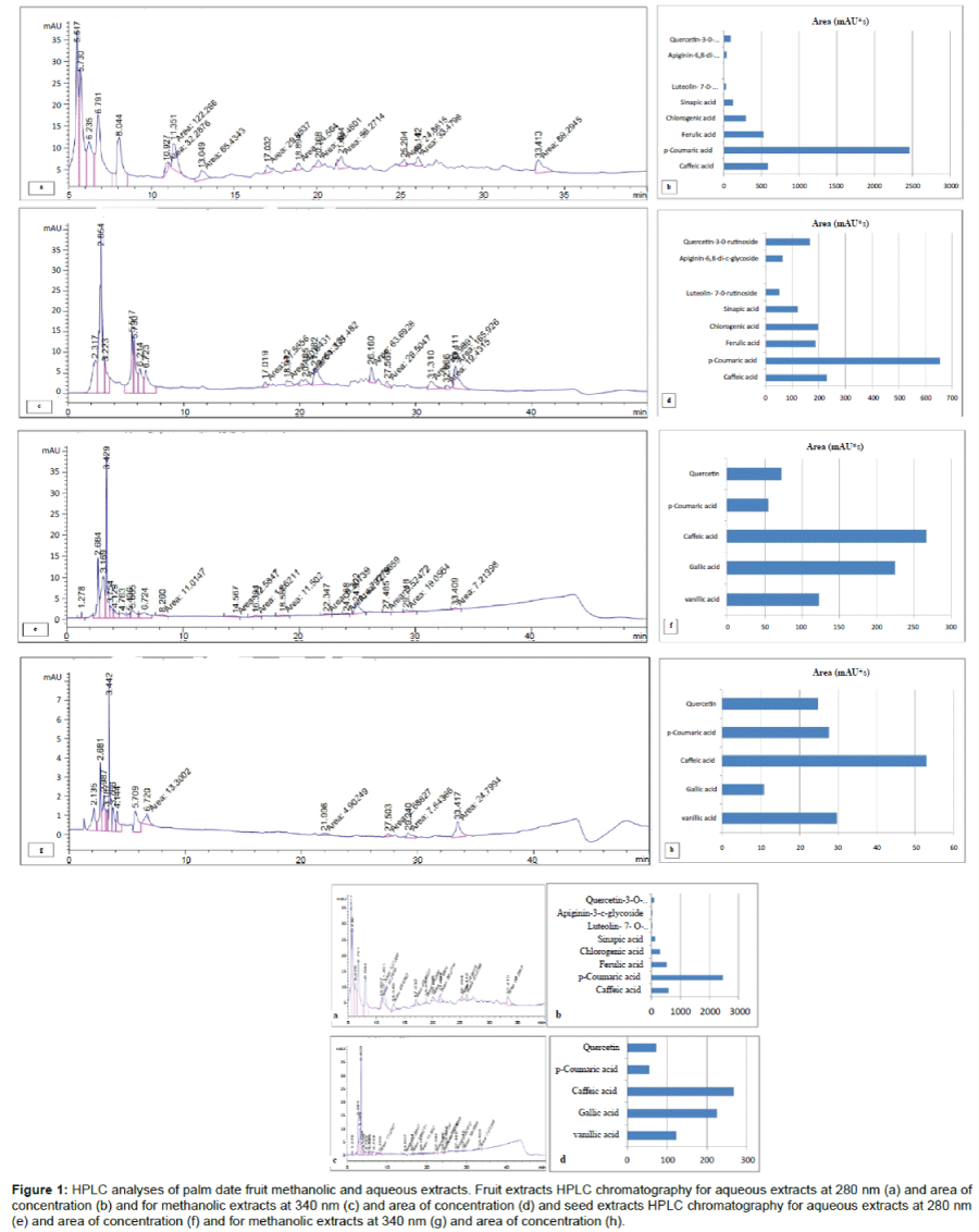
Gummy yellowish extract with a concentration of 40 g/100 g fruits was yielded from fruit extracts, while palm seed yielded dark brown extract with final concentration of 1.5 g/100 g. Eight compounds were detected by HPLC analysis for fruit methanolic and aqueous extracts. The compounds, from high to low concentration, were p-coumaric acid, caffeic acid, ferulic acid, chlorogenic acid, sinapic acid, quercitin- 3-o-glycoside, apiginin-c-glycoside and luteoline -7-o-glycoside respectively (Figures 1a and 1b). On other hand, five compounds were detected by HPLC analysis for seed methanolic and aqueous extracts. From high to low concentration, they were caffeic acid, gallic acid, vanillic acid, quercitin and p-coumaric acid respectively (Figures 1c and 1d). Moreover, the concentration of these substances in aqueous extracts was greater than in methanolic extracts in both fruits and seeds (Figures 1b and 1d).
Figure 1: HPLC analyses of palm date fruit methanolic and aqueous extracts. Fruit extracts HPLC chromatography for aqueous extracts at 280 nm (a) and area of concentration (b) and for methanolic extracts at 340 nm (c) and area of concentration (d) and seed extracts HPLC chromatography for aqueous extracts at 280 nm (e) and area of concentration (f) and for methanolic extracts at 340 nm (g) and area of concentration (h).
Results of animal survival and body weight in different groups
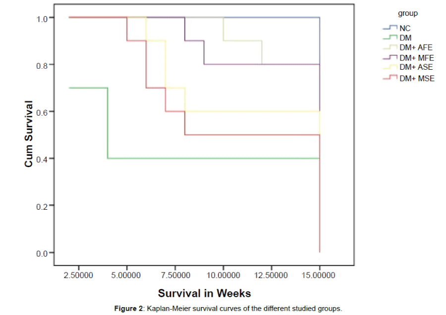
By the end of experiment 10 rats were survived in NC group, 4 rats in DM group, 8 rats in DM+AFE and DM+MFE groups, 6 rats in DM+ASE group and 5 rats in DM+MSE group. Meyer-Kaplan curve showed that survival rate was greater in aqueous extracts than methanolic extracts and in fruits than seed extracts (Figure 2). DC group showed significant increase in body weight compared to NC group (p<0.05). Also, compared to diabetic group, DM+AFE, DM+MFE, DM+ASE and DM+MSE group showed significant reduction in body weight (p<0.05). Moreover, we did not found any statistical significant difference in body weight among these groups (Table 1).
| NC group (n=10) |
DM group (n=4) |
DM+AFE group (n=8) |
DM+MFE group (n=8) |
DM+ASE group (n=6) |
DM+MSE group (n=5) |
|
|---|---|---|---|---|---|---|
| Body weight (g) | 176.93 ± 17.1 | 248.34 ± 15.9* | 194.60 ± 27.2# | 196.29 ± 10.5# | 189.86 ± 7.2# | 196.57 ± 15.5# |
| Fasting blood glucose (mg/dl) | 86.28 ± 6.77 | 464.71 ± 43.96* | 125.00 ± 13.6# | 154.71 ± 17.4*# | 173.14 ± 13.71*#$ | 197.71 ± 9.44*#$& |
| Serum cholesterol (mg/dl) | 135.14 ± 18.31 | 218.7 ± 25.52* | 147.46 ± 18.63# | 140.2 ± 10.19# | 141.2 ± 11.2# | 133.1 ± 16.0#$ |
| Serum triglycerides (mg/dl) | 102.5 ± 11.32 | 177.8 ± 22.6* | 122.8 ± 17.7# | 112.1 ± 17.2# | 109.5 ± 13.8# | 105.5 ± 11.5# |
| Serum HDL (mg/dl) | 51.0 ± 4.0 | 31.28 ± 9.8* | 41.5 ± 6.6 | 45.2 ± 7.47 | 47.4 ± 7.27 | 41.9 ± 13.2 |
| Serum LDL (mg/dl) | 63.6 ± 10.8 | 151.2 ± 13.77* | 81.3 ± 10.6# | 72.5 ± 5.92# | 74.4 ± 19.2# | 63.3 ± 6.7#$ |
All results are expressed as mean ± SD. One way ANOVA with Scheffe’s post hoc test (significance at p ≤ 0.05).*significant vs. NC group, #significant vs. DM group, $significant vs. DM+AFE group and significant vs. DM+MFE and €significant vs. group treated with ASE. NC: Normal Control, DC: Diabetic Control, DM+AFE: diabetic rats treated with aqueous Fruit extract, DM+MFE: diabetic rats treated with methanolic fruit extract, DM+ASE: diabetic rats treated with aqueous seed extract, DM+MSE: diabetic rats treated with methanolic seed extract.
Table 1: Body weight (g), fasting blood glucose (mg/dl) and Lipid profile (serum cholesterol (mg/dl), serum triglycerides (mg/dl), serum HDL (mg/dl) and serum LDL (mg/dl)) in different studied groups.
Results of fasting blood glucose (mg/dl) and lipid profile (serum TC, TGs, LDL and HDL) in different groups
DC group showed significant increase in fasting blood glucose compared to NC group (p<0.001) and treatment with seed and fruit extracts caused significant decrease in fasting blood glucose compared to diabetic group (p<0.05). Also, fruit extracts (AFE, MFE) groups showed more significant decrease in fasting blood glucose compared to seed extracts (ASE, MSE) (p<0.05) and aqueous extracts showed more significant reduction in fasting blood glucose than methanolic extracts (p<0.05) (Table 1).
DM group showed significant increase in serum TGs, TC and LDL with significant decrease in serum HDL compared to NC group (p<0.05) and this significant elevation was significantly attenuated in treated groups (AFE, MFE, ASE and MSE) compared to DM group (p<0.05). There were no statistical significant difference among all treated groups except in TC between AFE group and MSE group (p<0.05). On the other hand, HDL showed significant decrease in DM group compared to NC group and this attenuation was improved in all treated groups (p<0.05) (Table 1).
Results of markers of kidney functions (serum creatinine, urea, uric acid and urinary albumin excretion) in different groups
Diabetic rats showed significant increase in serum creatinine, urea and uric acid and urinary albumin excretion (UAE) compared to normal rats (p<0.05) and these parameters significantly attenuated in all treated groups (DM+AFE, DM+MFE, DM+ASE and DM+MSE) compared to DM groups (p<0.05). However, the degree of attenuation of these parameters was significantly higher in fruit extract groups (AFE and MFE) than seed extract groups (AFE and MFE) (p<0.05) and in aqueous extracts (AFE and ASE) groups than methanolic extract (MFE and MSE) groups (p<0.05) (Table 2).
| Serum creatinine (mg/dl) |
Serum urea (mg/dl) |
Serum uric acid (mg/dl) |
Urinary albumin excretion (UAE) (mg/24 h) |
|
|---|---|---|---|---|
| NC group (n=10) | 0.58 ± 0.066 | 23.71 ± 3.14 | 3.37 ± 0.13 | 19.57 ± 1.71 |
| DM group (n=4) | 2.92 ± 0.23* | 71.57 ± 2.14* | 6.67 ± 0.20* | 398.4 ± 14.19* |
| DM+AFE group (n=8) | 1.44 ± 0.097*# | 34.14 ± 3.71*# | 4.1 ± 0.14*# | 125.7 ± 5.52*# |
| DM+MFE group (n=8) | 1.92 ± 0.17*#$ | 50.57 ± 3.69*#$ | 4.82 ± 0.26*#$ | 282.2 ± 11.35*#$ |
| DM+ASE group (n=6) | 2.34 ± 0.09*#$& | 57.42 ± 1.51*#$& | 5.47 ± 0.19*#$& | 307.10 ± 13.8*#$& |
| DM+MSE group (n=5) | 2.77 ± 0.11*$&€ | 65.0 ± 1.63*#$&€ | 6.21 ± 0.13*#$&€ | 336.14 ± 7.92*#$&€ |
All results are expressed as mean ± SD. One way ANOVA with Scheffe’s post hoc test (significance at p ≤ 0.05).*significant vs. NC group, #significant vs. DM group, $significant vs. DM+AFE group and significant vs. DM+MFE and €significant vs. group treated with ASE. NC: Normal Control, DC: Diabetic Control, DM+AFE: diabetic rats treated with aqueous Fruit extract, DM+MFE: diabetic rats treated with methanolic fruit extract, DM+ASE: diabetic rats treated with aqueous seed extract, DM+MSE: diabetic rats treated with methanolic seed extract.
Table 2: Kidney function parameters (serum creatinine (mg/dl), urea (mg/dl), uric acid (mg/dl) and urinary albumin excretion (UAE) (mg/24 h)) in different studied groups.
Results of oxidative stress markers (MDA, GSH, SOD and catalase enzyme activities) in kidney tissues in different groups
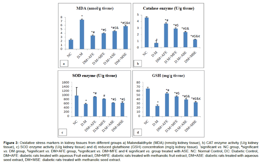
Kidney tissues obtained from diabetic rats showed significant increase in MDA concentration compared to those obtained from normal rats (p<0.001) (Figure 3a), while the activities of catalase and SOD enzymes as well as the concentration of GSH showed significant decrease in kidney tissues obtained from diabetic rats compared to those obtained from normal rats (p<0.001) (Figures 3b- 3d). These parameters showed significant improvement in all treated groups (DM+AFE, DM+MFE, DM+ASE and DM+MSE) compared to DM groups (p<0.05). However, the degree of improvement was significantly higher in fruit extract groups (AFE and MFE) than seed extract groups (ASE and MSE) (p<0.05) and in aqueous extracts (AFE and ASE) groups than methanolic extract (MFE and MSE) groups (p<0.05).
Figure 3: Oxidative stress markers in kidney tissues from different groups a) Malondialdhyde (MDA) (nmol/g kidney tissue), b) CAT enzyme activity (U/g kidney tissue), c) SOD enzyme activity (U/g kidney tissue) and d) reduced glutathione (GSH) concentration (mg/g kidney tissue). *significant vs. NC group, #significant vs. DM group, $significant vs. DM+AFE group, &significant vs. DM+MFE and € significant vs. group treated with ASE. NC: Normal Control, DC: Diabetic Control, DM+AFE: diabetic rats treated with aqueous Fruit extract, DM+MFE: diabetic rats treated with methanolic fruit extract, DM+ASE: diabetic rats treated with aqueous seed extract, DM+MSE: diabetic rats treated with methanolic seed extract.
Results of histopathological damage score in different groups
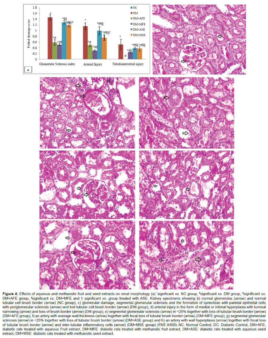
The damage score showed significant increase in glomerular sclerosis, arterial lesions and tubulointerstitial sclerosis in diabetic rats compared to normal rats (p<0.001) (Figure 4a). The degree of renal damage was significantly improved in all treated groups (DM+AFE, DM+MFE, DM+ASE and DM+MSE) compared to DM groups (p<0.001). However, the degree of improvement was significantly higher in fruit extract groups (AFE and MFE) than seed extract groups (AFE and MFE) (p<0.001), in AFE group than MFE group and in MSE group than ASE group (p<0.05). Kidneys obtained from NC group showed normal glomerular and tubular structure with normal vascular architecture (Figure 4b), while kidney obtained from DM group showed segmental glomerular sclerosis with formation of synechiae with parietal epithelial cells and periglomerular sclerosis, lost tubular cell brush border and arterial injury in the form of medial or intimal hyperplasia with luminal narrowing (Figures 4c and 4d), the degree of glomerular sclerosis and tubular injury was reduced in all treated groups (Figures 4e-4h).
Figure 4: Effects of aqueous and methanolic fruit and seed extracts on renal morphology (a) *significant vs. NC group, #significant vs. DM group, $significant vs. DM+AFE group, §significant vs. DM+MFE and † significant vs. group treated with ASE. Kidney specimens showing b) normal glomerulus (arrow) and normal tubular cell brush border (arrow) (NC group), c) glomerular damage, segmental glomerular sclerosis and the formation of synechiae with parietal epithelial cells with periglomerular sclerosis (arrow) and lost tubular cell brush border (arrow) (DM group), d) arterial injury in the form of medial or intimal hyperplasia with luminal narrowing (arrow) and loss of brush border (arrow) (DM group), e) segmental glomerular sclerosis (arrow) in <25% together with loss of tubular brush border (arrow) (DM+AFE group), f) an artery with average wall thickness (arrow) together with focal loss of tubular brush border (arrow) (DM+MFE group), g) segmental glomerular sclerosis (arrow) in <25% together with loss of tubular brush border (arrow) (DM+ASE group) and h) an artery with wall hyperplasia (arrow) together with focal loss of tubular brush border (arrow) and inter-tubular inflammatory cells (arrow) (DM+MSE group) (PAS X400). NC: Normal Control, DC: Diabetic Control, DM+AFE: diabetic rats treated with aqueous Fruit extract, DM+MFE: diabetic rats treated with methanolic fruit extract, DM+ASE: diabetic rats treated with aqueous seed extract, DM+MSE: diabetic rats treated with methanolic seed extract.
Discussion
The main findings of the present study can be summarized as follow a) diabetic nephropathy in type 2 DM was associated with dyslipidemia, impairment of kidney functions and morphology and enhanced redox state in kidney tissues b) pretreatment with either fruit (aqueous or methanolic) extracts or seed (aqueous or methanolic) extracts caused improvement in dyslipidemia, kidney functions and morphology and oxidative stress state in kidney tissue and c) fruit extracts offer powerful protective effects than seed extracts and aqueous showed more protective effects than seed extracts.
The present study demonstrated that type 2 DM was characterized by significant increase in body weight, fasting blood sugar, serum TC, TGs, LDL with significant decrease in animal survival and serum HDL. Previous studies reported similar findings such as Wang et al. [25], Khanra et al. [26] and Srimaroeng et al. [27]. Activation of hormone sensitive lipase in DM (normally inhibited by insulin) which increases the mobilization of free fatty acids from the peripheral fat deposits to blood stream might explain hyperlipidemia demonstrated in the present study. The elevated levels of serum lipids in DM cause the risk of development of diabetic nephropathy [28,29]. The significant increase in animal survival, body weight in type 2 diabetic rats in the present study might be due to the consumption of a diet rich in energy (saturated fats) and its deposition in different body fat stores [30] and reduction in energy expenditure [31]. On other hand, treatment with fruit and seed aqueous and methanolic extracts caused significant improvement in animal survival, body weight, fasting blood sugar, serum TC, TG, LDL and HDL. In agreement with these findings, Zangiabadi et al. [22] demonstrated that aqueous date extract improved body weight and fasting blood glucose in rat model of diabetic peripheral neuropathy. These findings suggest that consumption of palm date fruit and seed extracts could improve the body weight, hyperglycemia and hyperlipidemia in type 2 diabetes mellitus. Improvement in body weight in palm date extracts treated groups might be a reflection for improvement of hyperglycemia and hyperlipidemia.
Development of diabetic nephropathy in the present study was evidenced by significant increase in serum creatinine, uric acid, urea and urinary albumin excretion (UAE). In consistence with these findings, previous studies demonstrated significant increase in plasma creatinine and BUN [32-34] and serum urea levels [35] in diabetic rats compared to normal. The elevation of serum creatinine, uric acid, urea and UAE are reflection to impairment of kidney functions with development of diabetic nephropathy and loss of glomerular membrane permeability [36]. Histopathopathological examination for kidneys obtained from diabetic rats revealed significant increase in glomerular damage (segmental glomerular sclerosis and the formation of synechiae with parietal epithelial cells with periglomerular sclerosis), loss of tubular epithelial cell brush border and arterial injury in the form of medial or intimal hyperplasia with luminal narrowing. In agreement with these findings Khanra et al. [26] demonstrated thickening of glomerular and tubular basement membrane of kidneys in type 2 diabetic rats and Tervaert et al. [37] demonstrated mesangial expansion in type 2 diabetic rat’s kidney which might influence membrane permeability.
Treatment with fruit and seed aqueous and methanolic extracts caused significant improvement in kidney functions and morphology as evidenced by significant decrease in serum creatinine, urea, uric acid and urinary albumin excretion as well as significant reduction in glomerular and tubulointerstitial damage scores compared to diabetic untreated rats. These findings suggest renoprotective effects for fruit and seed aqueous and methanolic extracts against diabetic nephropathy in rats. Moreover, the effects of fruit extracts were more powerful than seed extracts and aqueous extracts were more powerful than methanolic extracts. This could be explained by the difference in compounds present in each type of extract as well as the concentration these compounds in the extracts. In the present study, fruit extracts yielded 8 compounds (p-coumaric acid, caffeic acid and ferulic acid have the highest concentration) while seed extracts yielded 5 compounds (caffeic acid and gallic acid have the highest concentration). Also, the concentrations of these compounds in both extracts were great in aqueous extracts than methanolic extracts. In line with agreement with El Arem et al. [18] attributed the nephroprotective effect of the aqueous extracts of palm date against the renal damage induced by dichloroacetic acid to its richness in antioxidant compounds such as ferulic, caffeic and p-coumaric acids.
Improvement of renal functions and morphology by palm dates in the present study might be due to control of hyperglycemia and hyperlipidemia in this animal model and due to the compounds present in palm date extracts such as quercitin and p-coumaric acid. In consistence with this hypothesis, Mallikarjun et al. [38] demonstrated that quercetin reduced the excretion of urinary albumin excretion by 40% at the 4th week in diabetic rats. In consistence with the findings of the present study, El Arem et al. [18] demonstrated that aqueous date extract (ADE) significantly improved the plasma levels of creatinine, urea and uric acid as well as kidney morphology in a rat model of DCA-induced nephropathy. Also, Al-Qarawi et al. [39] reported that administration of the extracts of the flesh and pits of palm date caused significant improvement in serum creatinine and urea concentrations and the proximal tubular damage in a gentamicin-induced nephrotoxicity in rats. Moreover, Saafi-Ben Salah et al. [17] showed that aqueous extract of Deglet Nour cultivar has improved serum creatinine and urea and attenuated the histopathological damage induced by dimethoate in kidney of rats. Also, Ali and Abdu [40] showed that the aqueous date extract of Ajwa cultivar has improved the histopathological change induced by Ochratoxin (A) in renal distal and proximal tubules.
Reactive oxygen species (ROS) and oxidative stress play a crucial role in diabetic pathophysiology [41] and play important role in high glucose-induced renal injury [42,43]. The present study demonstrated enhanced redox status within the kidney tissues of type 2 diabetic rats. This is evidenced by significant increase in MDA (product and marker of peroxidations of cell membrane lipids) with significant depletion in the endogenous antioxidants such as GSH, SOD and catalase enzyme activities in kidney tissues of diabetic untreated rats. Treatment with fruit and seed aqueous and methanolic extracts caused significant reduction in MDA in kidney tissues with significant increase in the endogenous antioxidants such as GSH, SOD and catalase enzymes activities. Moreover, the effect of aqueous fruit extract was more powerful compared to other types of extracts. Previous studies reported similar findings for palm date extracts. For instance, in a rat model of trichloroacetic acid (TCA)-induced hepatoxicity, El Arem et al. [16] demonstrated that aqueous palm fruit extract attenuated enhanced redox state in liver as evidenced by significant reduction in hepatic lipid peroxidation products (MDA) with improvement of antioxidant enzyme e.g. catalase, SOD and glutathione peroxidase activities. Al- Qarawi et al. [39] suggested that the nephroprotective action of extracts of the flesh and pits of Phoenix dactylifera in gentamicin-induced nephrotoxicity attributed to the antioxidant components of date such as melatonin, vitamin E and ascorbic acid. Also, Saafi-Ben Salah et al. [17] showed that date aqueous extract of Deglet Nour cultivar has attenuated lipid peroxidation and improved antioxidant defense system in dimethoate-induced nephrotoxicity. The protective effect of aqueous date extract to their high amounts of antioxidant substances such as polyphenols, trace elements, and vitamin C [13,44-46].
Conclusion
We demonstrated in the present study renoprotective effects for palm date fruit and seeds aqueous and methanolic extracts against diabetic nephropathy in rats of type 2 DM. Moreover, the nephroprotective effects of fruit extracts were more powerful than seed extracts. This nephroprotective action might be due to improvement of glucose and lipid homeostasis and the antioxidants properties of its contents such as p-coumaric acid, ferulic acid, caffeic, quercetin and chlorogenic acid.
Conflict of Interest
All authors declare that there was no conflict of interest.
References
- American Diabetes Association (2001) Diabetes: Vital statistics. Alexandra, VA: ADA.
- FaragYM, Al Wakeel JS (2011) Diabetic nephropathy in the Arab Gulf countries. Nephron ClinPract 119: c317-322.
- Jian WX, Peng WH, Jin J, Chen XR, Fang WJ, et al. (2012) Association between serum fibroblast growth factor 21 and diabetic nephropathy. Metabolism 61:853-859.
- Hovind P, Tarnow L, Rossing K, Rossing P, Eising S, et al. (2003) Decreasing incidence of severe diabetic microangiopathy in type 1 diabetes. Diabetes Care 26: 1258-1264.
- Yokoyama H, Okudaira M, Otani T, Sato A, Miura J, et al. (2000) Higher incidence of diabetic nephropathy in type 2 than in type 1 diabetes in early-onset diabetes in Japan. Kidney Int 58:302–311
- Lee HB, Yu MR, Yang Y, Jiang Z, Ha H (2003) Reactive oxygen species-regulated signaling pathways in diabetic nephropathy. J Am SocNephrol 14: S241-245.
- Yamagishi S, Matsui T (2010) Advanced glycation end products, oxidative stress and diabetic nephropathy. Oxid Med Cell Longev 3: 101-108.
- Yamagishi S, Imaizumi T (2005) Diabetic vascular complications: Pathophysiology, biochemical basis and potential therapeutic strategy. Curr Pharm Des 11:2279-2299
- Vayalil PK (2012) Date fruits (Phoenix dactylifera Linn): an emerging medicinal food. Crit Rev Food SciNutr 52: 249-271.
- Ahmed MB, Hasona NA, Selemain HA (2008) Protective Effects of Extract from Dates (Phoenix dactylifera L.) and Ascorbic Acid on Thioacetamide-Induced Hepatotoxicity in Rats. Iran J Pharma Res 7: 193-201
- Hussein AS, Alhadami GA, Khalil YH (1998) The use of dates and date pits in broilers starter and finishers diets. BioresourTechol 66: 219-223
- Vinson JA1, Zubik L, Bose P, Samman N, Proch J (2005) Dried fruits: excellent in vitro and in vivo antioxidants. J Am CollNutr 24: 44-50.
- Al-Farsi M, Alasalvar C, Morris A, Baron M, Shahidi F (2005) Comparison of antioxidant activity, anthocyanins, carotenoids, and phenolics of three native fresh and sun-dried date(Phoenix dactyliferaL.) varieties grown in Oman. J Agric Food Chem 53:7592–7599.
- Ragab AR, Elkablawy MA, Sheik BY, Baraka HN (2013) Antioxidant and tissue-protective studies on Ajwa extract: Dates from Al Madinah Al-Monwarah, Saudi Arabia. J Environ Anal Toxicol 3:1.
- Saafi EB, Louedi M, Zakhama AF, Najjard MF, Hammami M, et al. (2011) Protective effect of date palm fruit extract (Phoenix dactyliferaL.) on dimethoate induced-oxidative stress in rat liver. ExpToxicolPathol 63:433–441.
- El Arem A, Saafi EB, Ghrairi F, Thouri A, Zekri M, et al. (2014a) Aqueous date fruit extract protects against lipid peroxidation and improves antioxidant status in the liver of rats sub-chronically exposed to trichloroacetic acid. J PhysiolBiochem 70:451-464.
- Saafi-Ben Salah EB, El Arem A, Louedi M, Saoudi M, Elfeki A, et al. (2012) Antioxidant-rich date palm fruit extract inhibits oxidative stress and nephrotoxicity induced by dimethoate in rat. J PhysiolBiochem 68:47-58
- El Arem A, Thouri A, Zekri M, Saafi EB, Ghrairi F, et al. (2014) Nephroprotective effect of date fruit extract against dichloroacetic acid exposure in adult rats. Food ChemToxicol 65: 177-184.
- Hussein AM, El-Mousalamy AMD, Hussein SAM, Mahmoud SA (2015) Effects of Palm Dates (Phoenix dactylifera L.) Extracts on Hepatic Dysfunctions in Type 2 Diabetic Rat Model. WJPPS 4: 62-79.
- Pujari RR, Vyawahare NS, Kagathara VG (2011) Evaluation of antioxidant and neuroprotective effect of date palm (Phoenix dactyliferaL.) against bilateral common carotid artery occlusion in rats. Ind J ExpBiol 49: 627-633.
- Kalantaripour TP, Asadi-Shekaari M, Basiri M, Gholaamhosseinian NA (2012) Cerebroprotective effect of date seed extract (Phoenix dactylifera) on focal cerebral ischemia in male rats. J BiolSci 12: 180-185.
- Zangiabadi N, Asadi-Shekaari M, Sheibani V, Jafari M, Shabani M, et al. (2011) Date fruit extract is a neuroprotective agent in diabetic peripheral neuropathy in streptozotocin-induced diabetic rats: A multimodal analysis. Oxid Med Cell Longev 976948.
- Uehara Y, Hirawa N, Numabe A, Kawabata Y, Ikeda T, et al. (1994) Long-term infusion of kallikrein attenuates renal injury in Dahl salt-sensitive rats. Hypertension 24: 770–778.
- Nangaku M, Izuhara Y, Usuda N, Inagi R, Shibata T, et al. (2005) In a type 2 diabetic nephropathy rat model, the improvement of obesity by a low calorie diet reduces oxidative/carbonyl stress and prevents diabetic nephropathy.Nephrol Dial Transplant 20:2661-2669.
- Wang Y, Campbell T, Perry B, Beaurepaire C, Qin L (2011) Hypoglycemic and insulin-sensitizing effects of berberine in high-fat diet- and streptozotocin-induced diabetic rats. Metabolism 60: 298-305.
- Khanra R, Dewanjee S, K Dua T, Sahu R, Gangopadhyay M, et al. (2015) AbromaaugustaL. (Malvaceae) leaf extract attenuates diabetes induced nephropathy and cardiomyopathy via inhibition of oxidative stress and inflammatory response. J Transl Med 13: 6.
- Srimaroeng C, Ontawong A, Saowakon N, Vivithanaporn P, Pongchaidecha A, et al. (2015) antidiabetic and renoprotective effects of Cladophoraglomeratakützing extract in experimental type 2 diabetic rats: A potential nutraceutical product for diabetic nephropathy. J Diabetes Res 320167.
- Vaziri ND (2006) Dyslipidemia of chronic renal failure: the nature, mechanisms and potential consequences. Am J Physiol Renal Physiol 290: F262-272.
- Sugano M, Yamato H, Hayashi T, Ochiai H, Kakuchi J, et al. (2006) High-fat diet in low-dose-streptozotocin-treated hemi-nephrectomized rats induces all features of human type 2 diabetic nephropathy: A new rat model of diabetic nephropathy.NutrMetabCardiovasc Dis 16:477-484.
- Srinivasan K, Patole PS, Kaul CL, Ramarao P (2004) Reversal of glucose intolerance by pioglitazone in high fat diet-fed rats. Methods Find ExpClinPharmacol 26: 327-333.
- Storlien LH, James DE, Burleigh KM, Chisholm DJ, Kraegen EW (1986) Fat feeding causes widespread in vivo insulin resistance, decreased energy expenditure and obesity in rats. Am J Physiol 251: E576-583.
- Ha H, Yoon SJ, Kim KH (1994) High glucose can induce lipid peroxidation in the isolated rat glomeruli. Kidney Int 46: 1620-1626.
- Kang N, Alexander G, Park JK, Maasch C, Buchwalow I, et al. (1999) Differential expression of protein kinase C isoforms in streptozotocin-induced diabetic rats. Kidney Int 56: 1737-1750.
- King GL, Loeken MR (2004) Hyperglycemia-induced oxidative stress in diabetic complications. Histochem Cell Biol 122:333-338.
- Dewanjee S, Maiti A, Sahu R, Dua TK, Mandal V (2011) Effective control of type 2 diabetes through antioxidant defense by edible fruits of Diospyrosperegrina. Evi Based Compliment Alter Med 675397.
- Jensen PK, Christiansen JS, Steven K, Parving HH (1981) Renal function in streptozotocin-diabetic rats. Diabetologia 21: 409-414.
- Tervaert TWC, Mooyaart AL, Amann K, Cohen AH, Cook HT, et al. (2010) Renal pathology society. Pathologic classification of diabetic nephropathy. J Am SocNephrol 21:556–563.
- Mallikarjun BC, Smitha B, Shylaja MD, Rajan MGR, Salimath PV (2012) Development of a sensitive and specific immunoassay to evaluate diabetic nephropathy and dietary modulation in experimental animals. J Pharm Res 5: 3613-3617.
- Al-Qarawi AA, Abdel-Rahman H, Mousa HM, Ali BH, El-Mougy SA (2008) Nephroprotective action of Phoenix dactylifera. in gentamicin-induced nephrotoxicity. Pharm Biol 4:227–2230.
- Ali A, Abdu S (2011) Antioxidant protection against pathological mycotoxins alterations on proximal tubules in rat kidney. Func Foods Heals Dis 4: 118–134.
- Dewanjee S, Das AK, Sahu R, Gangopadhyay M (2009) Antidiabetic activity of Diospyrosperegrina fruit: Effect on hyperglycemia, hyperlipidemia and augmented oxidative stress in experimental type 2 diabetes. Food ChemToxicol 47:2679-2685.
- Ha H, Lee HB (2000) Reactive oxygen species as glucose signaling molecules in mesangial cells cultured under high glucose. Kidney IntSuppl 77: S19-25.
- Iglesias-De La Cruz MC, Ruiz-Torres P, Alcamí J, Díez-Marqués L, Ortega-Velázquez R, et al.(2001) Hydrogen peroxide increases extracellular matrix mRNA through TGF-ß in human mesangial cells. Kidney Int 59:87-95.
- Mansouri A, Embarek G, Kokkalou E, Kefalas P (2005) Phenolic profile and antioxidant activity of the Algerian ripe date palm fruit (Phoenix dactylifera). Food Chem 89: 411–420.
- Hong YJ, Tomas-Barberan FA, Kader AA, Mitchell AE (2006) The flavonoid glycosides and procyanidin composition of Deglet Noor dates (Phoenix dactylifera). J Agric Food Chem 54: 2405-2411.
- Allaith AAA (2008) Antioxidant activity of Bahraini date palm (Phoenix dactylifera L.) fruit of various cultiva rs. Int J Food SciTechnol 43: 1033-1040.
Relevant Topics
- Analytical Biochemistry
- Applied Biochemistry
- Carbohydrate Biochemistry
- Cellular Biochemistry
- Clinical_Biochemistry
- Comparative Biochemistry
- Environmental Biochemistry
- Forensic Biochemistry
- Lipid Biochemistry
- Medical_Biochemistry
- Metabolomics
- Nutritional Biochemistry
- Pesticide Biochemistry
- Process Biochemistry
- Protein_Biochemistry
- Single-Cell Biochemistry
- Soil_Biochemistry
Recommended Journals
- Biosensor Journals
- Cellular Biology Journal
- Journal of Biochemistry and Microbial Toxicology
- Journal of Biochemistry and Cell Biology
- Journal of Biological and Medical Sciences
- Journal of Cell Biology & Immunology
- Journal of Cellular and Molecular Pharmacology
- Journal of Chemical Biology & Therapeutics
- Journal of Phytochemicistry And Biochemistry
Article Tools
Article Usage
- Total views: 15681
- [From(publication date):
June-2016 - Aug 24, 2025] - Breakdown by view type
- HTML page views : 14296
- PDF downloads : 1385


