Make the best use of Scientific Research and information from our 700+ peer reviewed, Open Access Journals that operates with the help of 50,000+ Editorial Board Members and esteemed reviewers and 1000+ Scientific associations in Medical, Clinical, Pharmaceutical, Engineering, Technology and Management Fields.
Meet Inspiring Speakers and Experts at our 3000+ Global Conferenceseries Events with over 600+ Conferences, 1200+ Symposiums and 1200+ Workshops on Medical, Pharma, Engineering, Science, Technology and Business
Case Report Open Access
Crohn’s Disease-Small Bowel Obstruction Secondary to Enterolith-A Case Report
| Shah A1, Husainy MA1, Retnasingham G1, Burli P3 and Botchu R2* | |
| 1Department of Clinical Radiology, Leicester Royal Infirmary, Infirmary Square, Leicester LE1 5WW, UK | |
| 2Department of Clinical Radiology, Kettering General Hospital, Rothwell Road, Kettering, Northamptonshire NN16 8UZ, UK | |
| 3Department of Clinical Radiology, Lincoln County Hospital, Greetwell Road, Lincoln, Lincolnshire LN2 4AX, UK | |
| Corresponding Author : | Dr. Rajesh Botchu 11 Jackson way Kettering, UK, NN15 7DL Tel: +44 300 303 1573 E-mail: drrajeshb@gmail.com |
| Received April 01, 2013; Accepted April 20, 2013; Published April 25, 2013 | |
| Citation: Shah A, Husainy MA, Retnasingham G, Burli P, Botchu R (2013) Crohn’s Disease-Small Bowel Obstruction Secondary to Enterolith-A Case Report. OMICS J Radiology 2:119. doi: 10.4172/2167-7964.1000119 | |
| Copyright: © 2013 Shah A, et al. This is an open-access article distributed under the terms of the Creative Commons Attribution License, which permits unrestricted use, distribution, and reproduction in any medium, provided the original author and source are credited. | |
Visit for more related articles at Journal of Radiology
Abstract
Enteroliths (faecalith) are not uncommonly seen in inflammatory bowel disease especially Crohn’s disease. Small bowel obstruction secondary to enterolith in Crohn’s disease is extremely rare. We describe a case of small bowel obstruction secondary to enterolith formation which was successfully managed surgically. We also review the relevant literature.
| Keywords |
| Enterolith; Crohn’s disease; Small bowel obstruction |
| Introduction |
| It is estimated that nearly 2.2 million people in Europe have Crohn’s Disease (CD) [1]. Numerous intestinal and extraintestinal manifestations have been reported. The commonest cause for small bowel obstruction in CD is secondary to stricture. Enterolith formation associated with CD is a rare clinical entity with only 25 reported cases in 2005 and to the best of our knowledge only a further few cases reported since [2-7]. We report a 64 year old female with CD who presented with signs and symptoms of intestinal obstruction and was found to have small bowel obstruction due to an enterolith. |
| Case Report |
| A 64-year female presented to emergency department with 3 days history of right upper quadrant pain radiating to umbilicus with bilious vomiting. She was apyrexial and her observations were stable. Her blood biochemistry, including white cell count, was normal. She had a history of CD, diagnosed more than 20 years ago and had a previous right hemi-colectomy due to the disease. Other relevant past medical history included epilepsy that was well controlled with medications. |
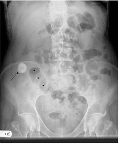
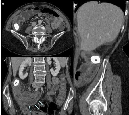
| Plain radiograph of the abdomen revealed a large calculus in the right iliac fossa with dilated loops of small bowel (Figure 1). A provisional diagnosis of gallstone ileus was made. An ultrasound of abdomen did not reveal any calculus within the gall bladder apart from biliary sludge. The known right iliac fossa calculus was again noted. To evaluate these further a CT with contrast of abdomen and pelvis was performed. This revealed a 3 cm lamellated calculus within thickened small bowel loops in the right iliac fossa causing proximal small bowel obstruction (Figure 2). No gallbladder calculus or evidence of cholecysto-duodenal fistula was demonstrated. Midline laparotomy was performed and the calculus was removed through an enterotomy, the inflammatory stricture was excised and an end-to-end anastomosis was performed. The patient made an uneventful recovery. |
| Discussion |
| CD is an inflammatory bowel disease (IBD) with transmural inflammation of bowel wall. The exact aetiology is unknown. CD has been shown to a have predominance for adolescent and young adults. It can involve any part of gastrointestinal tract, from the mouth to the anal canal. There is a predilection for the terminal ileum, accounting for a third of CD cases with a further third involving both the large and small bowel. |
| Enteroliths, which are endogenous foreign bodies and may result from agglutination of indigestible foreign materials. Less commonly, enteroliths form as a result of stasis, which promotes precipitation and concentration of the normal intestinal constituents. These are uncommon in the small bowel. A number of causes have been reported which promote intestinal stasis such as with Meckel’s diverticulum, small bowel diverticulosis, small bowel anastomosis and hypomotility. |
| Enteroliths in CD is rarely seen with about 30 reported cases. These are thought to be associated with patients with a long history of inflammatory bowel disease. The pathogenesis is thought to be due to intestinal fluid stasis secondary to strictures and malabsorption. Usually enteroliths are relatively small and pass into the colon without causing any symptoms. When there is an underlying stricture, the enterolith becomes obstructed and the stasis leads to further concentration of minerals increasing the size of the enterolith, making it less likely to pass. Large enteroliths may present with small bowel obstruction which can be complicated by perforation due to enterolith erosion. The formation of an enterolith in a Crohn’s patient is an important clue to a possible underlying pathology such as stricture formation. Stricturing CD occurs in 12–54% of the CD patient population and is associated with significant morbidity and can be associated with adenocarcinoma [8]. The risk of developing cancer increases after a disease duration of more than 10 years [9]. |
| The main differential for a radio-opaque density in the abdomen with small bowel obstruction is gallstone ileus, which is by far more common and calcified mesenteric adenopathy. |
| Reported case studies of small bowel enteroliths agree that surgical resection is the most appropriate management for these patients. A number of techniques have been described for the surgical removal of enteroliths either by manually crushing the stones and milking the fragments or removal through an enterotomy Due to the association of stricture formation and adenocarcinoma, surgical resection of the involved segment should be considered. This is particularly important in CD patients who are at increased risk and as it is often impossible to differentiate radiologically or macroscopically a benign stricture from a neoplastic one Therefore, surgical removal and resection of the stricture should be considered in CD patients presenting with enterolith obstruction. We have presented a case of CD with a long disease duration, who presented with signs and symptoms of intestinal obstruction due to an enterolith. One should be aware of this entity and include it in the differentials of small bowel obstruction in CD. |
References |
|
Figures at a glance
 |
 |
| Figure 1 | Figure 2 |
Post your comment
Relevant Topics
- Abdominal Radiology
- AI in Radiology
- Breast Imaging
- Cardiovascular Radiology
- Chest Radiology
- Clinical Radiology
- CT Imaging
- Diagnostic Radiology
- Emergency Radiology
- Fluoroscopy Radiology
- General Radiology
- Genitourinary Radiology
- Interventional Radiology Techniques
- Mammography
- Minimal Invasive surgery
- Musculoskeletal Radiology
- Neuroradiology
- Neuroradiology Advances
- Oral and Maxillofacial Radiology
- Radiography
- Radiology Imaging
- Surgical Radiology
- Tele Radiology
- Therapeutic Radiology
Recommended Journals
Article Tools
Article Usage
- Total views: 15688
- [From(publication date):
June-2013 - Aug 23, 2025] - Breakdown by view type
- HTML page views : 11024
- PDF downloads : 4664
