Research Article Open Access
Scytovirin Engineering Improves Carbohydrate Affinity and HIV-1 Entry Inhibition
| Hana McFeeters1, Morgan J. Gilbert1, Alexandra M. Wood1, Charity B. Haggenmaker1, Jennifer Jones2, Olaf Kutsch2 and Robert L. McFeeters1* | |
| 1Department of Chemistry, University of Alabama in Huntsville, 301 Sparkman Dr, Huntsville, AL 35899, USA | |
| 2Department of Medicine, Division of Infectious Diseases, University of Alabama at Birmingham, Birmingham, AL 35294, USA | |
| *Corresponding Author : | Robert L. McFeeters Department of Chemistry University of Alabama in Huntsville 301 Sparkman Dr, Huntsville AL 35899, USA Tel: (+1)-256-824-6023 Fax: (+1)-256-824-6349 Email: robert.mcfeeters@uah.edu |
| Received December 13, 2012; Accepted January 15, 2013; Published January 21, 2013 | |
| Citation: McFeeters H, Gilbert MJ, Wood AM, Haggenmaker CB, Jones J, et al. (2013) Scytovirin Engineering Improves Carbohydrate Affinity and HIV-1 Entry Inhibition. Biochem Physiol S2:003. doi:10.4172/2168-9652.S2-003 | |
| Copyright: © 2013 McFeeters H, et al. This is an open-access article distributed under the terms of the Creative Commons Attribution License, which permits unrestricted use, distribution, and reproduction in any medium, provided the original author and source are credited. | |
Visit for more related articles at Biochemistry & Physiology: Open Access
Abstract
Scytovirin, a cyanobacterium derived carbohydrate binding protein, acts as a potent HIV-1 entry inhibitor and could hold promise as a potential topical microbicide. Viral specificity is achieved as Scytovirin recognizes carbohydrate moieties rarely found in the extracellular matrix, but which are abundant on viral proteins. With the goal to improve the anti-viral capacity of Scytovirin, we here analyze the factors contributing to the Scytovirin anti-viral effect. We show that aromatic substitutions in the lower affinity C-terminal domain of Scytovirin lead to tighter carbohydrate binding. Several other mutations or an addition to the N-terminal abolish carbohydrate binding and abrogate the antiviral effect. Moreover, the increased binding affinity translates directly to improved antiviral efficacy. These studies improve our understanding of the Scytovirin:carbohydrate interaction and provide a blueprint for additional targeted mutations to advance Scytovirin as an entry inhibitor.
| Keywords |
| Scytovirin; Lectin; NMR; Antiviral entry inhibitor; Topical microbicide; Antiviral efficacy |
| Abbreviations |
| RT: Reverse Transcriptase; HIV: Human Immunodeficiency Virus; Gp: Glycoprotein; Man: Mannose; GlcNAc: N-acetylglucosamine; HSQC: Heteronuclear Single Quantum Coherence |
| Introduction |
| The continued emergence of multi-resistant forms of HIV-1 has driven the development of novel antiretrovirals over the years. Following the initial success of RT inhibitors and protease inhibitors, several classes of fusion or entry inhibitors were developed, some of which were transferred to clinical application. Acting before cellular infection, entry inhibitors have the advantage of being used as topical microbicides to prevent transmission. To date, several entry inhibitors targeting carbohydrates on the viral coat have been described. A variety of lectins isolated from plants, algae, prokaryotes, or invertebrate organisms have shown potential as topical microbicides [1-6]. These lectins bind to high-mannose moieties on the viral coat glycoproteins gp41 and gp120. Such high-mannose moieties are generally absent from cellular surface proteins. Inhibition on the order of nM has been reported [7] with strong preference for high-mannose carbohydrates and low affinity for the di or tri-mannose building blocks accounting for specificity of inhibition. |
| Scytovirin, a lectin with potent antiviral activity, comes from the cyanobacterium Scytonema varium [2]. The native protein has been characterized and recombinant expression and purification yielded protein with antiviral activity identical to that derived from native sources [2,8]. Scytovirin is a 10 kDa monomer, comprised of two nearly identical 39 amino acid segments (Figure 1). The repeat segments correspond to distinct structural domains that both bind carbohydrate [9]. The specificity of the interaction between Scytovirin and mannose containing oligosaccharides from HIV-1 coat glycoproteins was studied using a carbohydrate microarray chip [10]. Scytovirin showed high specificity towards high mannose moieties found on HIV envelope glycoproteins gp41 and gp120. High specificity for (Man)9 GlcNAc)2, henceforth referred to as Man9, was reported, with particular affinity to the D3 arm. |
| The affinity of Scytovirin for the D3 arm of Man9, Manα(1-2)Manα(1- 2)Manα(1-6) henceforth referred to as Man4, has been determined with the two nearly identical domains of Scytovirin having 7-fold different affinities [9]. These carbohydrate binding studies identified several regions involved in Man4 binding. Included in the C-terminal domain were residues Y54, W56, D57, and E58. Amino acids in this region are of interest for several reasons. First, the residue D57 is one of the three residues that are different between the two domains of Scytovirin and the only amino acid difference involved in carbohydrate binding. Second, the residues Y54 and W56 are in close proximity suggesting the importance of aromatic residues in carbohydrate binding. Also, the same aromatic-X-aromatic motif was previously found to play a central role in carbohydrate binding for Hevein and Hevein-like lectins [11,12]. The small size and high cysteine content (approximately 40 amino acids, 10 of which are cysteines) of Hevein carbohydrate binding domains are directly analogous to the carbohydrate binding domains of Scytovirin (39 amino acids, 8 of which are cysteines). |
| Guided by the possible implications of these previous findings, herein we report findings on Scytovirin mutants Y54F, W56L, D57N, E58D, and F85L. The mutants were recombinantly expressed and their structural integrity verified using heteronuclear NMR. Chemical shift perturbation mapping from a series of 15N-HSQC spectra acquired from titrations of isotopically labeled Scytovirin with HIV-1 like carbohydrate were recorded. Dissociation constants were obtained by fitting the changes in chemical shift to known concentrations of Man4 and Scytovirin. Affinity data were then correlated with functional HIV- 1 entry inhibition. Overall, a correlation between carbohydrate binding affinity and anti-HIV-1 efficacy was demonstrated. The results provide a blueprint for how Scytovirin potency for HIV-1 inhibition can be increased by protein engineering, resulting in stronger candidates for clinical trials. |
| Materials and Methods |
| Site directed mutagenesis |
| The gene for Scytovirin was commercially synthesized (GenScript, Piscataway NJ) using E. coli codon optimization and cloned into a pET32c vector using the NdeI and XhoI restriction sites. The N-terminal methionine was removed by site directed mutagenesis. The QuickChange Lightning kit (Agilent Technologies, Inc., Santa Clara, USA) was used to introduce mutations of interest into the Nand C-terminal domains. Since the two domains of Scytovirin are near sequence repeats, and there is consequently a high degree of nucleotide sequence identity, the strategy used to introduce mutations was a combination of site directed mutagenesis, restriction enzyme digest, and ligation. |
| In addition to the KpnI site in the multicloning site of pET32c, Scytovirin DNA contains a KpnI restriction site in the linker region of the protein that could be used to separate the N and C-terminal domains. For the DNA construct containing only the C-terminal domain, pET32c with Scytovirin DNA was digested by KpnI. The larger fragment of DNA containing the desired domain was isolated and religated. The ligation product was used for introduction of mutants using PCR based site directed mutagenesis. For the N-terminal domain, a silent mutation (GGT → GGG) was introduced to knock out the KpnI restriction site in the vector DNA (KpnI KO DNA). The KpnI site between the two sequence repeats of Scytovirin was preserved. After an XbaI/KpnI digest of the KpnI KO DNA, the smaller DNA fragment containing the N-terminal segment was isolated. Simultaneously, pET42b vector DNA was digested by XbaI/KpnI, the larger digestion product was isolated and used for ligation with the fragment containing the N-terminal domain of Scytovirin. For mutations in the N-terminal domain, mutant DNA was digested by XbaI/KpnI and smaller fragment was isolated. This fragment was ligated with the larger fragment resulting from XbaI/ KpnI digest of KpnI KO DNA yielding full-length Scytovirin with the desired mutation. Similarly for C-terminal domain mutants, pET32c DNA with the C-terminal domain mutation was digested by XbaI/KpnI and ligated with a smaller fragment of DNA from KpnI KO DNA XbaI/ KpnI digest. The correct DNA sequence for Scytovirin mutants was confirmed by DNA sequencing. |
| Expression and purification |
| Scytovirin mutants were expressed in Spectra 9 (Cambridge Isotope Laboratories, Andover MA) minimal 15N media for NMR spectroscopy. The protein was purified as previously described [9]. HPLC fractions containing Scytovirin were pooled and buffer exchanged into 20 mM MES, 100 μM EDTA, and 10 μM NaN3 as preservative, pH 5.5, the same buffer used for all NMR. |
| Oligosaccharide titrations |
| Concentrated 15N labeled samples of Scytovirin mutants were used for titration experiments with HIV-like oligosaccharide Man9, Manα(1-2)Manα(1-2)Manα(1-6). Chemical shift perturbations were measured from a series of 15N-HSQC spectra with increasing ratio of Man4: Scytovirin. Typically, 8 spectra were recorded with following Man4:Scytovirin concentration ratios–0:1, 0.25:1, 0.5:1, 1:1, 2:1, 4:1, 8:1, and 16:1. The starting protein concentration was 300 μM. The addition of Man4 had minimal effect on Scytovirin concentration (<5% change), though volume corrections were included for all Scytovirin concentrations in all analysis. |
| Dissociation constant determination |
| Chemical shift perturbation data served as input for dissociation constant determination using a two independent site model. All data were recorded on an 800 MHz Varian Inova spectrometer with room temperature probe. 15N-HSQC spectra were recorded with 1024 complex points in the direct dimension and 96 in the indirect dimension. Sweep widths were 15 ppm for 1H and 30 ppm for 15N. All peaks were processed with Gaussian windows and no linear prediction. Peaks were picked manually with center positions determined from fits to the entire peak. As previously published [9], carbohydrate binding affinity was determined from best fits of curves from chemical shift versus known concentrations of protein and carbohydrate using a modified procedure in Matlab (MathWorks, Natick MA, USA) kindly provided by Dr. David Fushman (University of Maryland, College Park, MD, USA). |
| Anti-HIV Assay |
| Samples of 15N Scytovirin and mutants were buffer exchanged into PBS, pH 7.4 and used in an assay to determine antiviral efficacies. Protein samples were serially diluted with PBS and final amounts used for the assay were 0 (negative control), 0.05, 0.1, 0.2, 0.41, 0.82, 1.63, and 3.25 μg/mL of protein. Different strains of HIV-1 clade B (NL4.3 (X4-tropic), CUCY (R5), WEAU (R5), TYBE (X4)) and wild type Scytovirin were incubated together for 30 minutes at 37°C prior to the addition of J2574-R5 cells [13]. For inhibition by mutants and thioredoxin fusion, only HIV-1 CUCY was used. After 48 hours, percentage of GFP fluorescence with respect to negative control (buffer only) was determined by Guava flow cytometer as described previously [14]. All experiments were run in duplicate. |
| Results |
| Structural integrity of Scytovirin mutants |
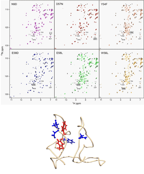
| NMR spectroscopy was used to evaluate the structural integrity of Scytovirin and all the mutants studied. Definitive of a well folded protein, the dispersion of peaks in the 15N-HSQC spectra was from 6.7 to 11.2 ppm in 1H and 100 to 133 ppm in 15N dimensions for wild type Scytovirin. Nearly identical chemical shift dispersion ranges were noted for all mutants studied. Also, a clear “resonance doubling” resulting from the almost identical amino acid sequences (Figure 1) and backbone fold of the N and C-terminal domains of Scytovirin has been reported [9]. Therefore, overlaying the wild type 15N-HSQC spectrum with the mutant spectra allowed for rapid determination of structural integrity and perturbations on a per residue basis. Chemical shift changes for all mutants are shown in supplemental figure 1 and those showing large changes listed in supplementary table 1. |
| 15N-HSQC spectra of the domain swap mutant D57N corresponded very well with the spectrum of wild type Scytovirin. See figure 2 for overlays of all mutant spectra with wild type. A reciprocal mutation in the N-terminal domain, N9D, also corresponded well with wild type. Only minor changes in chemical shifts of NH groups of residues close to the site of mutation were observed. The largest resonance shift was observed for either residue 9 or 57 in N9D or D57N, respectively, a result of the amino acid change, not a structural difference. 15N-HSQC spectra of aromatic Y54F, W56L, and F85L mutants showed somewhat larger changes in chemical shifts of NH groups in the proximity of the altered residue. Again, these changes are located in the proximity of the mutated site, expected from ring current effects. Overall, however, the spectra still correlated well with that of wild type Scytovirin. For the E58D mutant, the change in the C-terminal domain was much more pronounced. This relatively conservative mutation caused minor displacement for a majority of NH peaks in the C-terminal domain. Larger displacements were observed for NH groups of residues in immediate proximity of the mutated position. Most of the C-terminal peaks could not be assigned by inspection of the overlaid spectra. Further, if residue 58 was mutated into leucine, chemical shifts for C-terminal domain NH groups experienced even larger changes. From the 15N-HSQC of E58L it is evident that a majority of the C-terminal domain resonances do not overlay well with the wild type spectrum and a large number of peaks are observed near 8.3 ppm in 1H dimension. This is a strong sign of unfolded, random coil in the C-terminal domain. Therefore, no further studies of the E58L mutant were conducted. This also suggests that the domains of Scytovirin can fold separately, even with the shared disulfide bond. |
| Carbohydrate binding of Scytovirin mutants |
| The affinity of Scytovirin for high mannose carbohydrates common to HIV-1 envelope glycoproteins was determined from NMR titrations. A set of 15N-HSQC spectra of Scytovirin was recorded with increasing amounts of Man4. The amount of carbohydrate was correlated to the percent change in NH peak position (100% determined from complete saturation) and used to calculate the dissociation constant, Kd (Table 1). The analysis was performed using a two-site model of binding as previously described [9], except for the E58D mutation. Kd values for the mutants were compared to those of the wild type. To determine the effect of the only amino acid difference involved in carbohydrate binding, two “domain swapped” mutants were constructed. The D57N domain swapped mutant, in which the asparagine residue from the higher affinity N-terminal domain was substituted into the lower affinity C-terminal domain, did not increase the affinity of Scytovirin for Man4. In fact, the carbohydrate affinity decreased roughly 2-fold. Thus it appears the single residue difference between the N- and C-terminal carbohydrate binding domains is not responsible for the difference in affinity. As expected, a decrease in carbohydrate affinity was observed for the reverse mutation N9D. In both cases, the affinity worsened and Kd value increased to about 80 μM for the N-terminal domain and 580 μM for the C-terminal domain. |
| For the aromatic residue mutations, both Y54F and W56L mutants increased the affinity of Scytovirin for Man4. The Y54F mutant decreased the dissociation constant for the carbohydrate for the C-terminal domain from 250 to 75 μM. For the W56L mutant, the decrease in dissociation constant was on the same order, from 250 to 95 μM. Conversely, the F85L change significantly weakened carbohydrate binding, increasing the dissociation constant from 250 to 425 μM. Therefore it is possible to engineer Scytovirin to have higher affinity for the carbohydrate moieties on HIV-1 envelop glycoproteins. Future possibilities include analogous mutations in the N-terminal domain and potential additive effects from double mutants in the C-terminal domain (Y54F/W56L) or combined mutants in both domains. |
| Titration experiments for E58D with Man4 revealed that resonances in the C-terminal domain were not affected by Man4, even at high concentrations. Though not unambiguously assigned due to large chemical shift changes, no peaks outside of the N-terminal domain were affected by carbohydrate binding. Thus the C-terminal domain did not bind Man4, requiring single site binding analysis of the data. Residues in the N-terminal domain showed changes consistent with wild type Scytovirin, and a similar Kd. The unchanged affinity in the N-terminal domain suggests that the two carbohydrate binding sites of Scytovirin function independently with no cooperativity (i.e. a Hill coefficient equal to 1). |
| Anti-HIV-1 assays |
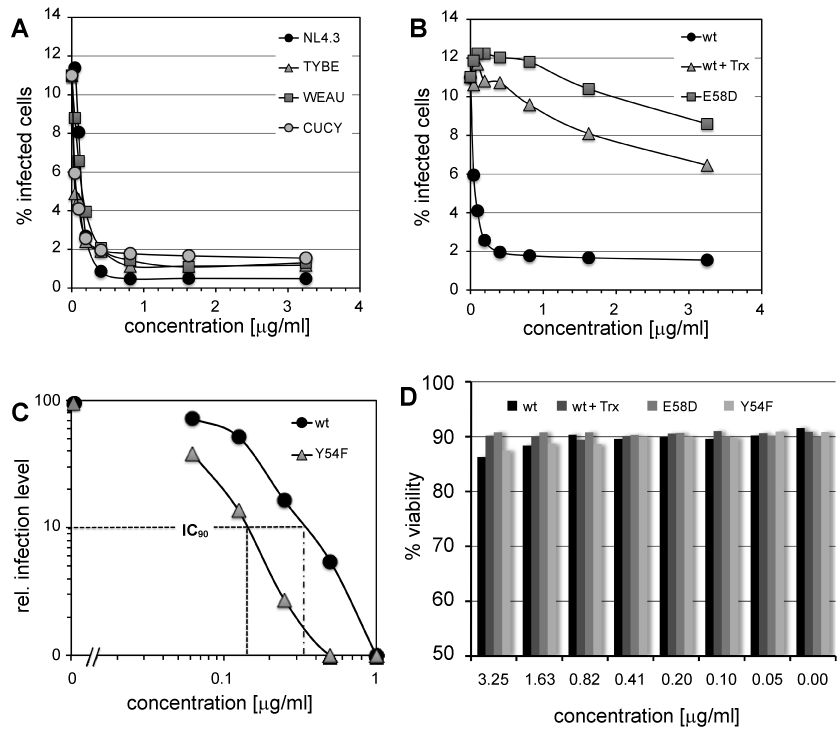
| Wild type Scytovirin and mutants were assayed for anti-HIV activity using a GFP reporter cell line, in which GFP expression serves as a surrogate marker of active HIV-1 infection [14]. In these infection assays, the obtained IC50 values were in the range of 6-18 nM for wild type Scytovirin, in good agreement with previous reported values [2,15]. Wild type Scytovirin was shown to inhibit both primary isolates and laboratory adapted HIV-1 strains. No differences were noticed for tropism, equally inhibiting X4, R5 or dual tropic strains (Figure 3A). No effect of Scytovirin (wild type, all single site mutants, and thioredoxin fusion) on cell viability at all concentrations studied was observed (Figure 3D). This indicates negligible cytotoxicity for Scytovirin. |
| To determine the effect of carbohydrate binding affinity, multivalency of binding, and N-terminal perturbations on antiviral efficacy, the higher affinity Y54F and W56L, and single site E58D mutants were tested in comparison to the wild type protein. The same dual tropic HIV-1 primary isolates were used for all assays. The high-affinity Y54F mutant was indeed more efficacious than wild type Scytovirin (Figure 3C), suggesting that carbohydrate binding affinity as determined by chemical shift perturbation mapping, is predictive of antiviral Scytovirin activity. The E58D mutant and loss of C-terminal domain carbohydrate binding did not show a high degree of HIV-1 inhibition even at concentrations where complete inhibition of wild type Scytovirin was observed (Figure 3B). An extrapolation led to an estimated IC50 near 1 μM, an 80-fold decrease. It thus appears that the two domains work together to achieve inhibition, and the higher affinity N-terminal domain is a more important determinant of efficacy. |
| The first two N-terminal residues of Scytovirin have been shown to play an important role in gp120/gp41 binding [16]. Therefore, we investigated effects of having the thioredoxin fusion tag still present with full-length wild type Scytovirin (as it is recombinantly expressed before protease cleavage during purification) [8] to determine the effect of N-terminal perturbation on inhibition of HIV-1 entry. Similar to the single carbohydrate binding site E58D mutant, the effect was very unfavorable on HIV-1 inhibition (Figure 3B). Even at high concentrations at which wild type Scytovirin inhibited HIV-1 infection, the thioredoxin fusion construct was only able to achieve 40% inhibition. The estimated IC50 from the extrapolated curve is roughly 2-fold lower than for E58D, yet clearly different than for a disruption of the C-terminal carbohydrate binding domain. The possibility of individual domains of Scytovirin as inhibitors follows. Previous reports indicate that the N and C-terminal domains by themselves have the same antiviral efficacy against HIV-1. If accurate, the implication is that a non-functional domain impairs the antiviral abilities. It also suggests that the tighter affinity carbohydrate binding mutations will significantly improve the antiviral efficacy of the single domains. |
| Discussion |
| Lectins that bind to carbohydrates on gp120/gp41 hold considerable promise as antiviral entry inhibitors. They act before infection, eliminating the necessity of the drugs crossing the cellular membrane. They also can be implemented as microbicides aimed at preventing sexual transmission of HIV. The principal potential of such inhibitory lectins is emphasized by the fact that multiple broadly neutralizing HIV- 1 antibodies target carbohydrate epitopes found on coat glycoproteins [17,18]. Further, glycan side chains are added by cellular mechanisms, which are not accessible to mutation induced by selective drug pressure. The virus could only escape the selective pressure of such lectin-based entry inhibitors by mutations that remove amino acids bearing glycan side chains. However, glycan side chains are essential for viral entry. In addition, it has been conclusively demonstrated that under the pressure of the humoral immune response, the virus evolves to actually add more glycan side chains to protect the gp120/gp41 complex from the binding of neutralizing antibodies (evolving glycan shield) [19]. Thus there is a continuous effort to develop lectins into therapeutically useful compounds. Scytovirin is unique among lectins in its high specificity for carbohydrate moieties found on HIV glycoproteins [10]. Such specificity may contribute to the lack of cellular toxicity. |
| Development of lectins as antiviral therapeutics for clinical use can be aided by protein engineering. Improving antiviral efficacy leads to decreased material per dose, which in turn reduces side effects, lowers the chance of an immune reaction, and reduces cost. Shorter constructs generally have reduced immunogenicity. Thus a minimal Scytovirin construct with maximum carbohydrate affinity (and thereby maximal antiviral efficacy) would be most ideal for viral entry inhibition. |
| Here we demonstrate that protein engineering of Scytovirin can improve carbohydrate binding affinity. Removing the hydroxyl group from the lower affinity C-terminal domain tyrosine by mutation to phenylalanine (Y54F) increased Scytovirin affinity for Man4. It thus appears that this aromatic side chain is intimately involved in the interaction with carbohydrate. However the data reported here showed that the aromatics of Scytovirin do not interact in the same fashion as those reported for Hevein-like proteins where the aromatic- X-aromatic motif is necessary for oligosaccharide binding [11,12]. Replacing the second aromatic with an aliphatic residue (W56L) also improved carbohydrate affinity. The third aromatic (F85) was also found to be critical for the interaction with carbohydrate, suggesting spatial proximity preserves the two aromatic interaction. It appears that aromatic interactions are important for Scytovirin carbohydrate binding, just not following the Hevein sequence motif. Further studies are required to determine if the specificity for Man4 changes with changes in aromatic composition, and if it impacts Scytovirin toxicity. So far, no toxicity has been observed in anti-viral assays. |
| The inability to bind (or exceedingly low affinity for) monosaccharide and extended region on Scytovirin showing changes upon carbohydrate binding [9] imply that Man4 has extensive contacts with Scytovirin. It seems that the specific monosaccharide linkages are key to recognition, not just interaction with aromatics residues of Scytovirin. Also, the one amino acid difference involved in carbohydrate binding (N9 in the N-terminal domain and D57 in the C-terminal domain) is not the major determinant of difference in binding affinity. Substitution of the high affinity, N-terminal domain asparagine into the equivalent position in the low affinity, C-terminal domain site diminished the interaction. Therefore a structural difference in the two domains must be responsible for the observed difference in carbohydrate binding affinities. Albeit a small structural change could account for the approximately 4 kJ/mol energetic differences (on the order of one hydrogen bond). |
| A correlation between binding affinity and antiviral efficacy was also observed. However the relationship appears to be more complicated than a simple, linear, one-to-one relation. Further illustrating the multivariable nature of the relationship, the loss of carbohydrate binding in a single domain for the full length protein was not equivalent to individual carbohydrate binding domains. Previous studies of the N-terminal domain alone showed efficacy against HIV-1 nearly indistinguishable for that of wild type Scytovirin [16]. The E58D mutant that resulted in a single carbohydrate binding site inhibited HIV-1 with markedly lower efficacy than wild type. Assuming accuracy/integrity in the single domain studies, it appears that the presence of the inactive C-terminal domain in full-length Scytovirin negatively impacts the anti-HIV-1 properties of the N-terminal domain. |
| Our results show that protein engineering can be used to improve Scytovirin as a therapeutic agent. Future studies of analogous N-terminal domain mutations are planned. Continued improvements in carbohydrate affinity and lowered immunogenicity will make Scytovirin a stronger antiviral entry inhibitor. |
| Acknowledgements |
| The authors thank Mary Hames for critical reading of the manuscript, Whitney Hedges for preliminary work on the Scytovirin NMR titration data, and the OVPR at UAHuntsville for URII financial support for this project. |
References
|
Tables and Figures at a glance
| Table 1 |
Figures at a glance
 |
 |
 |
| Figure 1 | Figure 2 | Figure 3 |
Relevant Topics
- Analytical Biochemistry
- Applied Biochemistry
- Carbohydrate Biochemistry
- Cellular Biochemistry
- Clinical_Biochemistry
- Comparative Biochemistry
- Environmental Biochemistry
- Forensic Biochemistry
- Lipid Biochemistry
- Medical_Biochemistry
- Metabolomics
- Nutritional Biochemistry
- Pesticide Biochemistry
- Process Biochemistry
- Protein_Biochemistry
- Single-Cell Biochemistry
- Soil_Biochemistry
Recommended Journals
- Biosensor Journals
- Cellular Biology Journal
- Journal of Biochemistry and Microbial Toxicology
- Journal of Biochemistry and Cell Biology
- Journal of Biological and Medical Sciences
- Journal of Cell Biology & Immunology
- Journal of Cellular and Molecular Pharmacology
- Journal of Chemical Biology & Therapeutics
- Journal of Phytochemicistry And Biochemistry
Article Tools
Article Usage
- Total views: 14387
- [From(publication date):
specialissue-2013 - Aug 16, 2025] - Breakdown by view type
- HTML page views : 9771
- PDF downloads : 4616
