Research Article Open Access
The Disc Positioning after Arthroscopic Lysis and Lavage of the Temporomandibular Joint
| Paulo Alexandre da Silva1*, Leonard Duarte Moreira2 and Fernando Silva Freire1 | |
| 1Department of Oral and Maxillofacial Surgery, Vivalle Hospital, São José dos Campos, SP, Brazil | |
| 2Department of Oral and Maxillofacial Surgery, Ouro Verde Hospital, São Leopoldo Mandic School of Dentistry, Cam-pinas, SP, Brazil | |
| Corresponding Author : | Paulo Alexandre da Silva Head of Oral and Maxillofacial Surgery Department of the Vivalle Hospital Av. 9 de Julho, 959 Jardim Apollo, São José dos Campos SP, Brazil, CEP (ZIP): 11 600-000, Brazil Tel: 55 12 98144 6000 E-mail: drpauloalexs@gmail.com |
| Received January 02, 2016; Accepted January 20, 2016; Published January 22, 2016 | |
| Citation: da Silva PA, Moreira LD, Freire FS (2016) The Disc Positioning After Arthroscopic Lysis and Lavage of the Tem-poromandibular Joint. J Pain Relief 5:224. doi:10.4172/2187-0846.1000224 | |
| Copyright: © 2016 da Silva PA, et al. This is an open-access article distributed under the terms of the Creative Commons Attribution License, which permits unrestricted use, distribution, and reproduction in any medium, provided the original author and source are credited. | |
Visit for more related articles at Journal of Pain & Relief
Abstract
The symptomatic disc displacement of the temporomandibular joint (TMJ) can be treated by many ways, like oclusal splints, physiotherapy, arthrocentesis, arthroscopy and open surgery. TMJ Arthroscopic Lysis and Lavage (ALL) has showed good results in these cases. The aimed of this study were to observe the disc positioning after the procedure and the improvement of pain and mouth opening. 38 (6 men and 32 women) with 54 symptomatic disc displacement were included in this study. The results showed improvement of the disc positioning in 37 TMJ (68.51%), mouth opening in 33 patients (86.84%) and pain 35 in patients 35 patients (92.7%). In conclusion ALL can improve the disc positioning, mouth opening and pain in patients with symptomatic TMJ disc displacement.
| Keywords |
| Arthroscopic lysis; Temporomandibular joint |
| Introduction |
| The internal derangement (DI) of the Temporomandibular joint (TMJ), specifically those with disc displacement can be treated by many different kinds, implying that conservative and reversible treatments, for example: intra-oral devices, physiotherapy and medicines and, in some cases, it need surgical treatments like arthrocentesis, arthroscopy and open surgeries, each on with its own rate of effectiveness. This study aimed to observe the disc position after TMJ Arthroscopic lysis and lavage (ALL) through post-procedural MRI and the success rate of pain reduction in jaw function and mouth opening. |
| Materials and Methods |
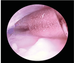
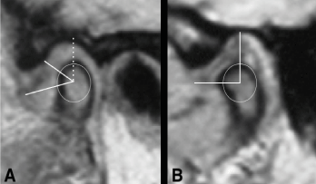
| The patients included in this study were 38 (6 men and 32 women), 54 TMJ, aged 16-55 years, with a mean age of 29.7 years old, treated between January 2013 and October 2014. These patients had limited mouth opening (MIO) >30 mm, pain in jaw function through visual analogic scale (VAS>5, positive for joint load test), magnetic resonance imaging (MRI) showing disc displacement: with reduction (DDWR) or without reduction (DDWoR). The TMJ were classified by Wilkes Stage [1]. All patients were refractory to conservative treatment. Comorbidities were investigated in laboratory (blood tests) according to the information obtained during the anamnesis in order to exclude the involvement of systemic disorders such as rheumatoid arthritis. Under general anesthesia, Arthroscopic lysis and lavage (ALL) as performed according to the puncture and triangulation technique described by McCain [2]. All the procedures were performed by the same surgeon using 1.9 mm zero-degree arthroscope, cannulas, sharp trocar, blunt obturator, knife, probe and bipolar electrode (Karl Storz Endoscopy, Tuttlingen-Germany) were used. Mechanical adhesion removal, disc mobilization and synovitis cauterization were performed under irrigation with saline solution (Figure 1). At the end of the procedure, 20 mg of sodium hyaluronate (Polireumin - TBR Pharma, São Paulo-Brazil) was used for infiltration in the upper joint space. The patients were advised to consume soft diet for 30 days, use a splint, and perform passive mouth-opening, mandible lateralization and protrusion during the first 15 days and after that they were referred to return to physiotherapy. Physiotherapy and splints were maintained for 3 months after surgery. All patients were evaluated during the postoperative period, specifically after 24 h, 72 h, 7 days, 15 days, 30 days, and beyond, for monthly assessments of the improvements of the pain in jaw function (VAS and load test) and the MIO. A control MRI was performed 6 months after ALL to assess the disc positioning and compare it to the initial MRI, overlap them (Figure 2) |
| Results |
| The patients presented with a mean MIO of 21.2 mm and TMJ pain with mean VAS of 7.85 and positive for joint load test.The Wilkes Stages found in the 54 TMJ (38 patients) were II-11 III-23, IV-16, V-4. Of the The mean MIO improvement index was 86.84% (33 patients - range: 62.7-96.8%), with the smallest and largest opening measurements of 36 mm and 52 mm, respectively, and a mean of 45 mm; the mean improvement index in pain in jaw function was 92.7% (35 patients - range: 71.9-95.3%), with low and high VAS of 0 and 4, respectively, and a mean of 2.3 (Table 1). Control MRI at 6 months post-surgery when compared with initial MRI showed: improvement disc positioning in 37 TMJ (68.51%): 8-Wilkes II, 17 Wilkes III, 12 Wilkes IV; the same disc positioning 13 TMJ (24.07%): 3 Wilkes II, 5 Wilkes III, 3 Wilkes IV and 2 Wilkes V; 4 TMJ (7.42%) worsening disc positioning: 1-Wilkes III, 1 Wilkes IV, 2 Wilkes V (Table 2). |
| Discussion |
| Previous studies have shown positive results for patients after TMJ Arthroscopy Lysis and Lavage (ALL) to improve the MIO and pain, being very stable at long-term [3]. The success rate of ALL for Murakami et al. [4], Friedrich et al. [5], Ohnuki et al. [6] were 91%, 82%, 74% respectively. But these results vary in the literature, suggesting that the success of ALL depends on the Wilkes Stage of the TMJ [7]. In this regard, Bronstein and Merrill [8] reported success rates of 96% for stage II, 83% for stage III, 88% for stage IV, and 63% for stage V. In addition, Smolka and Iizuka [9] described a mean success rate of 86.7%, which varied between 75% and 92.3% according to the TMJ stage. This variability was also noted in the current study and the means obtained were consistent with those in previously published studies with regard to pain reduction and an improved MIO, which were stable during the follow-up period. However, studies about disc positioning after ALL and its correlation with improvement or not of the MIO and pain in function are rare and there are not consensus in the literature to date. Ohnuki et al. [10] had been observed that just 10% of the discs displaced were repositioned after TMJ arthroscopy, and in cases of warped discs the results were worse in follow-up MRI, by the other way showed a improvement in mouth opening and painful symptoms concluding that, the disc function, disc mobility is more important than the disc position. Clark et al [11] using control MRI evaluations found a new disc positioning closer to the anatomical position, Silva et al. [12] showed at follow-up MRI a rate of 63% better disc position after ALL. Moses e Topper [13] concluded about the new disc position that was not related to disc repositioning, but instead to the mobilization and removal of the adhesions and degenerative inflammatory products. In this study was showed the improvement of anatomic relocation of the disc, but we also can see that in some cases the disc maintained the same position and another few cases your position worsened. Another important observation in this study was that the improvement of the disc positioning was not directly related to the improvement of MIO and pain. In some cases discs remained in the same position or their position worsened, but the patients improved MIO and pain, and other cases that the disc improved your position, patients maintained their initial symptoms. Therefore we agree that disc function (mobility) is more important than its position. The difficulties of the present study was to standardize the sample with only patients with disc displacement and pain and limited opening mouth who were refractory to conservative treatment, and the standardization of the Magnetic Resonance Image (MRI) as well. Further studies should investigate the issue correlating the disc positioning after ALL and its influence on MIO and pain in jaw function. |
| Conclusions |
| Arthroscopic Lysis and Lavage (ALL) can improve the articular disc positioning, MIO and reduced pain in jaw function, but the mobility of the disc is more important than its position. Further studies, including a long-term follow-up, are required to consolidate these results. |
References
- Wilkes CH (1989) Internal derangements of the temporomandibular joint. Pathological variations.Arch Otolaryngol Head Neck Surg 115: 469-477.
- McCain JP,Hossameldin RH (2011) Advanced arthroscopy of the temporomandibular joint.Atlas Oral Maxillofac Surg Clin North Am 19: 145-167.
- Leibur E,Jagur O, Müürsepp P, Veede L, Voog-Oras U (2010) Long-term evaluation of arthroscopic surgery with lysis and lavage of temporomandibular joint disorders.J Craniomaxillofac Surg 38: 615-620.
- Murakami K,Hosaka H, Moriya Y, Segami N, Iizuka T (1995) Short-term treatment outcome study for the management of temporomandibular joint closed lock. A comparison of arthrocentesis to nonsurgical therapy and arthroscopic lysis and lavage.Oral Surg Oral Med Oral Pathol Oral RadiolEndod 80: 253-257.
- Fridrich KL, Wise JM, Zeitler DL (1996) Prospective comparison of arthroscopy and arthrocentesis for temporomandibular joint disorders.J Oral Maxillofac Surg 54: 816-820.
- Ohnuki T, Fukuda M, Iino M, Takahashi T (2003) Magnetic resonance evaluation of the disk before and after arthr oscopic surgery for temporomandibular joint disorders.Oral Surg Oral Med Oral Pathol Oral RadiolEndod 96: 141-148.
- González-García R, Rodríguez-Campo FJ (2011) Arthroscopic lysis and lavage versus operative arthroscopy in the outcome of temporomandibular joint internal derangement: a comparative study based on Wilkes stages.J Oral Maxillofac Surg 69: 2513-2524.
- Bronstein SL, Merrill RG (1992) Clinical staging for TMJ internal derangement: application to arthroscopy. J CraniomandibDisord 6: 7-15.
- Smolka W,Iizuka T (2005) Arthroscopic lysis and lavage in different stages of internal derangement of the temporomandibular joint: correlation of preoperative staging to arthroscopic findings and treatment outcome.J Oral Maxillofac Surg 63: 471-478.
- Ohnuki T, Fukuda M, Nakata A, Nagai H, Takahashi T, et al. (2006) Evaluation of the position, mobility, and morphology of the disc by MRI before and after four different treatments for temporomandibular joint disorders.DentomaxillofacRadiol 35: 103-109.
- Clark G T, Sanders B, Bertolami C H (1993). Advances in diagnostic and surgical arthroscopy of the temporomandibular joint. Philadelphia: WB Saunders
- Silva PA, Lopes MT, Freire FS (2015) A prospective study of 138 arthroscopies of the temporomandibular joint.Braz J Otorhinolaryngol 81: 352-357.
- Moses JJ, Topper DC (1991) A functional approach to the treatment of temporomandibular joint internal derangement.J CraniomandibDisord 5: 19-27.
Tables and Figures at a glance
| Table 1 | Table 2 |
Figures at a glance
 |
 |
| Figure 1 | Figure 2 |
Relevant Topics
- Acupuncture
- Acute Pain
- Analgesics
- Anesthesia
- Arthroscopy
- Chronic Back Pain
- Chronic Pain
- Hypnosis
- Low Back Pain
- Meditation
- Musculoskeletal pain
- Natural Pain Relievers
- Nociceptive Pain
- Opioid
- Orthopedics
- Pain and Mental Health
- Pain killer drugs
- Pain Mechanisms and Pathophysiology
- Pain Medication
- Pain Medicine
- Pain Relief and Traditional Medicine
- Pain Sensation
- Pain Tolerance
- Post-Operative Pain
- Reaction to Pain
Recommended Journals
Article Tools
Article Usage
- Total views: 11164
- [From(publication date):
January-2016 - Aug 29, 2025] - Breakdown by view type
- HTML page views : 10162
- PDF downloads : 1002
