 Spanish
  Chinese
  Russian
  German
  French
  Japanese
  Portuguese
  Hindi
Hindi Our Group organises 3000+ Global Conferenceseries Events every year across USA, Europe & Asia with support from 1000 more scientific Societies and Publishes 700+ Open Access Journals which contains over 50000 eminent personalities, reputed scientists as editorial board members.
Open Access Journals gaining more Readers and Citations
700 Journals and 15,000,000 Readers Each Journal is getting 25,000+ Readers
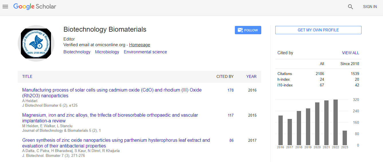
Google Scholar citation report
Citations : 3330
Journal of Biotechnology & Biomaterials received 3330 citations as per Google Scholar report
Indexed In
- Index Copernicus
- Google Scholar
- Sherpa Romeo
- Open J Gate
- Genamics JournalSeek
- Academic Keys
- ResearchBible
- China National Knowledge Infrastructure (CNKI)
- Access to Global Online Research in Agriculture (AGORA)
- Electronic Journals Library
- RefSeek
- Hamdard University
- EBSCO A-Z
- OCLC- WorldCat
- SWB online catalog
- Virtual Library of Biology (vifabio)
- Publons
- Geneva Foundation for Medical Education and Research
- Euro Pub
- ICMJE
Useful Links
Recommended Journals
Related Subjects
Share This Page
In Association with
Characterization of biomaterials using AFM based fast nanoscale imaging and quantitative nanomechanical techniques
2nd Annual Conference and Expo on BIOMATERIALS
T Neumann, T Muller, D Stamov, H Haschke, C Pettersson, S Kostrowski and T Jahnke
JPK Instruments AG, Germany
Posters & Accepted Abstracts: J Biotechnol Biomater
Abstract
Besides structural and physico-chemical composition, topography, roughness, adhesiveness as well as mechanical properties of biomaterials are the relevant factors making them suitable for biomedical applications. All these factors affect cell differentiation and tissue formation, and are crucial for their integration as well as healing capacity in the human body. Atomic Force Microscopy is suitable for measuring all of these characteristics with nanometer scale resolution under physiological conditions. We have developed a multipurpose AFM device allowing comprehensive characterization of biological samples such as live cells, tissues and biomaterials in the nanoscale. True optical integration allows the simultaneous use of advanced inverted optical microscope techniques such as DIC or confocal laser scanning microscopy, but also upright optics, such as macroscopes for the investigation of opaque samples. With our â�?�?Quantitative Imagingâ�? (QIâ�?¢) mode several sample properties, such as the topography, stiffness and adhesiveness, can be obtained with one measurement in high resolution. Even more complex data like Young�?´s modulus images, topography at different indentation forces in terms of tomography, or recognition events can be obtained. A variety of biological samples have been investigated to demonstrate the capability and flexibility of QIâ�?¢. The NanoWizard�?® ULTRA Speed technique allows fast AFM imaging of dynamic processes with approximately 1 frame per second. The kinetics of collagen type I fibrillogenesis was imaged in situ with high spatiotemporal resolution, revealing the formation of the 67 nm D-banding hallmark. With the CellHesion�?® technique, the adhesion of a single living cell to any substrate can be measured and validated using comprehensive analysis tools. The side-view cantilever holder enables a side view of the cell-sample interface while performing adhesion experiments, providing complementary information without expensive z-stacking. The inherent drawbacks of traditional AFM imaging modes for fast imaging or for challenging samples like living cells can be overcome by the NanoWizard�?® ULTRA Speed and QIâ�?¢ mode. We present an enhancement of the AFM technique providing a versatile tool for an extensive characterization of biomaterials.Biography
Email: confregis@jpk.com