 Spanish
  Chinese
  Russian
  German
  French
  Japanese
  Portuguese
  Hindi
Hindi Our Group organises 3000+ Global Conferenceseries Events every year across USA, Europe & Asia with support from 1000 more scientific Societies and Publishes 700+ Open Access Journals which contains over 50000 eminent personalities, reputed scientists as editorial board members.
Open Access Journals gaining more Readers and Citations
700 Journals and 15,000,000 Readers Each Journal is getting 25,000+ Readers
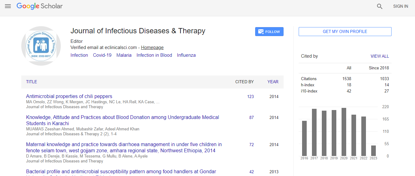
Google Scholar citation report
Citations : 1529
Journal of Infectious Diseases & Therapy received 1529 citations as per Google Scholar report
Indexed In
- Index Copernicus
- Google Scholar
- Open J Gate
- RefSeek
- Hamdard University
- EBSCO A-Z
- OCLC- WorldCat
- Publons
- Euro Pub
- ICMJE
Useful Links
Recommended Journals
Related Subjects
Share This Page
Immunohistochemistry approach in encephalitozoonosis
3rd Annual Congress on Infectious Diseases
Adriano Pereira and Maria Anete Lallo
S�?£o Paulo State University, Brazil
Posters & Accepted Abstracts: J Infect Dis Ther
Abstract
Encephalitozoonosis is a common disease of a wide range of mammalian hosts caused by Encephalitozoon cuniculi and the detection of this pathogen in tissue samples is considered difficult. Microsporidia spores of E. cuniculi can be observed in histological sections stained with routine dyes in tissues of experimentally infected animals in the laboratory because the amount of spores is generally large, but in veterinary clinics of domestic and wild animals these stains are often not sufficient for diagnosis. Then, for these cases a variety of techniques, including special staining methods, immunohistochemistry (IHC), electron microscopy and molecular methods are used for diagnosis and exclusion of other microorganisms. The aim of this study was to describe about the use of IHC for the detection of E. cuniculi in tissue samples that have been published to some groups of researchers and veterinary pathologists throughout the world. An English literature search was done through databases (MEDLINE; NCBI, Bethesda, MD, USA) in order to examine publications. We considered papers from 1993 to today that described IHC analysis performed using formalin fixed and paraffin-embedded tissue sections. The diagnosis of encephalitozoonosis using IHC has been made in rabbits (brain, kidneys, lungs, heart, liver, eyes, and spleen), dogs (eyes), horse (villi ofvallantochorion), South American fur seal (lungs, spleen and kidneys), squirrel monkey (brain), emperor tamarins (blood capillaries, arteries, heart, liver, lung, brain and Kidney), cats (brain and kidney), cotton-top tamarins (kidneys and blood), chicken (esophagus, intestine, liver, kidneys, heart, skeletal muscle and brain) and snow leopard (eyes). E. cuniculi was successfully identified in different kind of tissues using IHC. Based on our results, we suggest that IHC should be regarded as a useful tool both for specific demonstration of E. cuniculi and for its localization within tissues helping researchers and veterinary pathologists for the diagnosis of encephalitozoonosis.Biography
Adriano Pereira is a Teacher in the areas of health and biological sciences at São Camilo University, São Paulo, Brazil. He has done Master’s degree in Veterinary Medicine and PhD in Environmental and Experimental Pathology. His research involves studying microsporidia with a focus on biology and immune response against this emerging and opportunistic pathogen.