Case Report Open Access
Contrast-Enhancing Lesions within the Spinal Chord Suggests Immune Reconstitution Inflammatory Syndrome (IRIS) in a Patient with Natalizumab Associated Progressive Multifocal Leukencephalopathy (Natalizumab-PML)
| Justus Carl Marquetand*, Felix Bischof and Thomas Nägele | |
| Center for Neurology Hertie Institute for Clinical Brain Research Department Cellular Neurology, Germany | |
| Corresponding Author : | Justus Carl Marquetand Center for Neurology Hertie Institute for Clinical Brain Research Department Cellular Neurology Otfried-Müller-Straße 27 72076 Tubingen, Germany Tel: +497071/290 E-mail: justus.marquetand@med.uni-tuebingen.de |
| Received April 17, 2015; Accepted June 15, 2015; Published June 18, 2015 | |
| Citation: Marquetand JC, Bischof F, Nägele T (2015) Contrast-Enhancing Lesions within the Spinal Chord Suggests Immune Reconstitution Inflammatory Syndrome (IRIS) in a Patient with Natalizumab Associated Progressive Multifocal Leukencephalopathy (Natalizumab-PML). J Neuroinfect Dis S1:001. doi:10.4172/2314-7326.S1-001 | |
| Copyright: © 2015 Marquetand JC, et al. This is an open-access article distributed under the terms of the Creative Commons Attribution License, which permits unrestricted use, distribution, and reproduction in any medium, provided the original author and source are credited. | |
| Related article at Pubmed, Scholar Google | |
Visit for more related articles at Journal of Neuroinfectious Diseases
Abstract
Background: Many patients with natalizumab-associated progressive multifocal leukoencephalopathy (PML) who are treated by plasma exchange (PLEX) develop an immune reconstitution inflammatory syndrome (IRIS), which often leads to severe tissue destruction within the brain. While steroids can efficiently treat IRIS, they worsen PML and a reliable and prompt diagnosis of IRIS in natalizumab-PML patients is thus critical.
Clinical presentation: We report on a patient with supratentorial manifestations of natalizumab-PML after PLEX and corresponding new gadolinium-enhancing lesions within the spinal cord, which were clinically corresponding to difficulties to urinate.
Conclusion: Cerebral MRI supported the diagnosis of IRIS. Therefore, detection of contrast-enhancing lesions within the spinal cord may indicate IRIS in patients with natalizumab-PML.
| Keywords |
| Pml; Iris; Spinal; Natalizumab |
| Abbreviations |
| MS: Multiple Sclerosis; PML: Progressive Multifocal Leukencephalopathy; IRIS: Immune Reconstitution Inflammatory Syndrome; PLEX: Plasma Exchange; JCV: John-Cunnigham-Virus; MRI: Magnet Resonance Imaging; MRS: Magnet Resonance Proton Spectroscopy; NAA: N-Acetyl-Aspartate; CSF: Cerebrospinal Fluid; PCR: Polymerase Chain Reaction; CNS: Central Nervous System; HIV: Human Immunodeficiency Virus |
| Background |
| Progressive multifocal leukoencephalopathy (PML) is an opportunistic infection of the central nervous system (CNS) caused by reactivation of the papilloma JC-virus (JCV). Latent infection with JCV is common and reactivation occurs in individuals with reduced immunocompetence within the CNS (e.g. acquired immune deficiency syndrome (AIDS) [1] due to HIV-infection, after bone-marrow transplantation or under treatment with a growing number of immunosuppressive or immunomodulatory drugs. In patients with multiple sclerosis (MS), treatment with the monoclonal antibody natalizumab blocks 47-integrin on the surface of circulating lymphocytes and prevents their migration into the CNS. The reported risk of PML is about one in 500 MS patients treated with natalizumab for more than 24 months. Patients with natalizumab-PML are commonly treated with plasma exchange (PLEX) to remove natalizumab from the circulation. After a median of 32 days following PLEX [2], patients develop an immune reconstitution inflammatory syndrome (IRIS), which is characterized by severe inflammation within the brain that often leads to rapid and persistent tissue destruction [3]. IRIS is pathologically characterized by massive infiltration of CD8+ lymphocytes into the CNS and commonly accompanied by destruction of the blood-brain barrier, which can be identified by gadolinium enhancement in cerebral MRI [4]. To prevent IRIS-induced tissue destruction, patients are treated with high dose intravenous steroids [5]. Steroids have now been clearly shown to worsen PML [6] and a prompt and accurate diagnosis of IRIS in PML patients is thus critical. |
| Clinical Presentation |
| A 33-year-old female patient was admitted to our hospital with a two-weeks history of progressive right-sided hemiparesis. She was first diagnosed with relapsing-remitting MS at the age of 26, and initially treated with glatirameracetate for 2.5 years until she became pregnant and treatment was changed to intravenous immunoglobulin. Following delivery, MS disease activity was high and she was put on natalizumab, which was well tolerated. During this therapy, no new relapses occurred. Anti-JCV antibodies in serum were positive. Before presenting to our hospital, she had received 22 infusions of natalizumab within the preceding 27 months. |
| Initial examination revealed a right-sided spastic brachiofacial hemiparesis with increased reflexes, positive Babinski sign and myoclonus. She did not have aphasia or dysarthria, the other cranial nerves and sensitivity were intact as was bladder and bowel function. EDSS upon admission was 4.5, she was able to walk without aid and rest about 300m and acquired minimal assistance in meal preparation. |
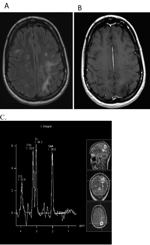
| Cerebral MRI including MR- proton spectroscopy (MRS) revealed several periventricular T2 hyperintense lesions and new patchy subcortical white matter hyperintensities in the left parietal lobe with faint contrast-enhancement (Figure 1). Long echo time MRS (TE=135ms) showed a discrete lactate signal at 1.3 ppm as a sign of inflammation together with a decrease of N-acetyl-aspartate (NAA). Choline was almost normal indicating that there were no signs of acute demyelination. Previous MRI of the spinal cord were unremarkable. |
| Motor evoked potentials to the right arm were absent and to the lower limbs were delayed, as were visually evoked potentials on the right side. EEG and ophthalmologic assessment were normal. A CSF analysis showed a normal cell count and protein concentration, increased local IgG-production and positive oligoclonal bands. CSF PCR for JC-Virus was positive with 50 copies / ml. |
| Treatment with 250 mg mefloquin per week and 45mg mirtazapine per day were initiated and five courses of PLEX were performed. During the following two weeks, her hemiparesis increased and she developed a sensorimotor aphasia and a hemianopsia. She experienced two tonic-clonic seizures and was put on levetiracetam. |
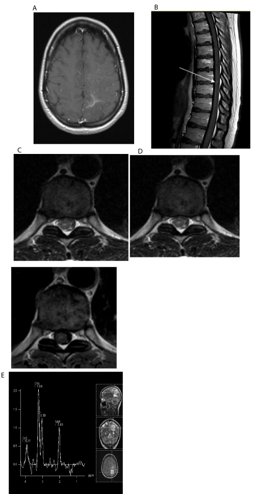
| Seventeen days following PLEX, she experienced difficulties to urinate and a spinal MRI revealed a corresponding contrast-enhancing lesion within the spinal cord (Figure 2 b-d). Cerebral MRI showed pronounced contrast-enhancement along the borders of the PML lesions and MRS showed a marked increase of choline (Cho) and lactate and a decrease of NAA indicating acute demyelination (Figure 2a). Collectively, these findings confirmed IRIS that presented with a symptomatic spinal manifestation. IRIS was treated with infusions of 2g methylprednisolone per day for five consecutive days, which led to resolution of the bladder problems. |
| Conclusion |
| Many patients with natalizumab-PML who are treated with PLEX develop IRIS [7]. The diagnosis of IRIS currently relies on the detection of rapidly evolving neurological symptoms and contrast-enhancing lesions or brain swelling in cerebral MRI [8,9] and proton magnetic resonance spectroscopy may add to the diagnosis [10,11]. |
| The disease course of this patient was in line with other reported cases (e.g. number of natalizumab infusision, etc. [3]. Here we demonstrated that new spinal cord symptoms and Gd+ lesions within the spinal cord could indicate the onset of IRIS in natalizumab-PML patients and thus add to the diagnosis. This may be particularly advantageous in patients in whom IRIS is difficult to differentiate from PML, as is the case in patients who display contrast-enhancing PML lesions [3,12] or patients who develop IRIS without contrast-enhancing cerebral lesions [9,13]. While IRIS is commonly considered to occur predominantly at sites of PML lesions [14], this case demonstrates that Gd+ IRIS lesions can occur in areas not affected by PML on conventional MRI including the spinal cord. |
| The management of PML and IRIS is becoming increasingly relevant due to the growing number of drugs associated with the development of PML as has recently been exemplified by the Janus kinase inhibitor Ruxolitinib [15,16]. The complexity of PML and IRIS is increased by other manifestations of JCV infection including granular cellular neuronopathy [17] or meningitis [18] and the development of new opportunities to treat PML including continuous infusion of interleukin- 7 [19] and immunization with recombinant particles of JCVproteins [19] and treatment of IRIS with the CCR5 antagonist maraviroc [20]. |
| Acknowledgement |
| We are grateful to our patient for her participation and her consent to publish these data. |
| Consent |
| Written informed consent for publication of the clinical details and clinical images was obtained from the patient. A copy of the consent form is available for review by the Editor of this journal. |
References
- Berger JR (2013) PML diagnos tic criteria: consensus statement from the AAN Neuroinfectious Disease Section. Neurology 80: 1430-1438.
- Dahlhaus S (2013) Disease course and outcome of 15 monocentrically treated natalizumab-associated progressive multifocal leukoencephalopathy patients.J NeurolNeurosurg Psychiatry 84: 1068-1074.
- Johnson T, ANath (2011) Immune reconstitution inflammatory syndrome and the central nervous system.CurrOpinNeurol 24: 284-290.
- Gray F (2005) Central nervous system immune reconstitution disease in acquired immunodeficiency syndrome patients receiving highly active antiretroviral treatment. J Neurovirol 11 Suppl 3: 16-22.
- Clifford DB (2010)Natalizumab-associated progressive multifocal leukoencephalopathy in patients with multiple sclerosis: lessons from 28 cases. Lancet Neurol 9: 438-446.
- Antoniol C (2012) Impairment of JCV-specific T-cell response by corticotherapy: effect on PML-IRIS management? Neurology 79: 2258-2264.
- Vermersch P (2011) Clinical outcomes of natalizumab-associated progressive multifocal leukoencephalopathy. Neurology 76: 1697-1704
- McCarthy M, Nath A (2010)Neurologic consequences of the immune reconstitution inflammatory syndrome (IRIS).CurrNeurolNeurosci Rep 10: 467-475.
- Martin-Blondel G (2013)In situ evidence of JC virus control by CD8+ T cells in PML-IRIS during HIV infection. Neurology 81: 964-970.
- Cuvinciuc V (2010)Proton MR spectroscopy of progressive multifocal leukoencephalopathy-immune reconstitution inflammatory syndrome. AJNR Am J Neuroradiol 31: E69-70.
- Gheuens S (2012)Metabolic profile of PML lesions in patients with and without IRIS: an observational study. Neurology 79: 1041-1048.
- Yousry TA (2012)Magnetic resonance imaging pattern in natalizumab-associated progressive multifocal leukoencephalopathy. Ann Neurol 72: 779-787.
- Tan K (2009)PML-IRIS in patients with HIV infection: clinical manifestations and treatment with steroids. Neurology 72: 1458-1464.
- Tan CS,Koralnik IJ (2010)Progressive multifocal leukoencephalopathy and other disorders caused by JC virus: clinical features and pathogenesis. Lancet Neurol 9: 425-437.
- Bloomgren G (2012)Risk of natalizumab-associated progressive multifocal leukoencephalopathy.N Engl J Med 366: 1870-1880.
- Wathes R (2013)Progressive multifocal leukoencephalopathy associated with ruxolitinib. N Engl J Med 369: 197-198.
- AgnihotriSP (2014)JCV GCN in a natalizumab-treated MS patient is associated with mutations of the VP1 capsid gene. Neurology 83: 727-732.
- Agnihotri SP (2014)A fatal case of JC virus meningitis presenting with hydrocephalus in a human immunodeficiency virus-seronegative patient. Ann Neurol 76: 140-147.
- Sospedra M (2014) Treating Progressive Multifocal LeukoencephalopathyWith Interleukin 7 and Vaccination With JC Virus Capsid Protein VP1. Clin Infect Dis 59: 1588-1592.
- Giacomini PS (2014)Maraviroc and JC virus-associated immune reconstitution inflammatory syndrome. N Engl J Med 370: 486-488.
Figures at a glance
 |
 |
| Figure 1 | Figure 2 |
Relevant Topics
- Bacteria Induced Neuropathies
- Blood-brain barrier
- Brain Infection
- Cerebral Spinal Fluid
- Encephalitis
- Fungal Infection
- Infectious Disease in Children
- Neuro-HIV and Bacterial Infection
- Neuro-Infections Induced Autoimmune Disorders
- Neurocystercercosis
- Neurocysticercosis
- Neuroepidemiology
- Neuroinfectious Agents
- Neuroinflammation
- Neurosyphilis
- Neurotropic viruses
- Neurovirology
- Rare Infectious Disease
- Toxoplasmosis
- Viral Infection
Recommended Journals
Article Tools
Article Usage
- Total views: 14613
- [From(publication date):
specialissue-2015 - Aug 24, 2025] - Breakdown by view type
- HTML page views : 10025
- PDF downloads : 4588
