Make the best use of Scientific Research and information from our 700+ peer reviewed, Open Access Journals that operates with the help of 50,000+ Editorial Board Members and esteemed reviewers and 1000+ Scientific associations in Medical, Clinical, Pharmaceutical, Engineering, Technology and Management Fields.
Meet Inspiring Speakers and Experts at our 3000+ Global Conferenceseries Events with over 600+ Conferences, 1200+ Symposiums and 1200+ Workshops on Medical, Pharma, Engineering, Science, Technology and Business
Image Open Access
Medical Image: Coronary Atherosclerosis
| Brandon Micsh* | |
| School of Health Sciences, University of Canterbury, New Zealand | |
| Corresponding Author : | Brandon Micsh School of Health Sciences, University of Canterbury New Zealand Tel: 64 3 326 2907 E-mail: brandonmicsh@gmail.com |
| Received: October 26, 2015 Accepted: October 27, 2015 Published: October 30, 2015 | |
| Citation: Micsh B (2015) Medical Image: Coronary Atherosclerosis . J Obes Weight Loss Ther 5:i002. doi: 10.4172/2165-7904.1000i002 | |
| Copyright: © 2015 Micsh B. This is an open-access article distributed under the terms of the Creative Commons Attribution License, which permits unrestricted use, distribution, and reproduction in any medium, provided the original author and source are credited. | |
Visit for more related articles at Journal of Obesity & Weight Loss Therapy
Medical Image
Coronary artery disease develops in a body when major blood vessels that supply heart with blood, oxygen and nutrients get damaged. Plaque (cholesterol containing deposits) in arteries and inflammation are the main factors for coronary artery disease.
Plaques build up narrows your coronary arteries and decrease blood flow to the heart. Finally, the decreased blood flow causes angina that is called chest pain, short breathing and other coronary artery disease signs and symptoms. A complete blockage due to plague causes a heart attack may result into death.
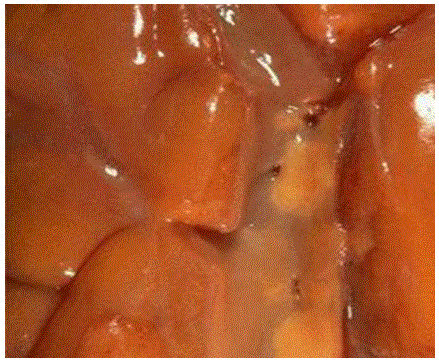
Below is the photograph is of a patient suffering with mild coronary atherosclerosis (Figure 1). A few scattered yellow lipid plaques can be seen on the intimal surface of the opened coronary artery traversing the epicardial surface of a heart. The degree of atherosclerosis here is not significant enough to cause disease, but could be the harbinger of worse atherosclerosis to come [1,2].
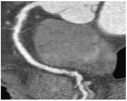
This is the Computed tomography angiogram of the same patient suffering from coronary atherosclerosis (Figure 2). (Upper Portion: Mild proximal stenosis with expansive remodelling and predominantly nonexpansive plaque; Lowe Portion: Partially calcified advanced mid to distal stenosis).
References
- http://library.med.utah.edu/WebPath/ATHHTML/ATH002.html
- http://www.clevelandclinicmeded.com/medicalpubs/diseasemanagement/cardiology/coronaryarterydisease/
Figures at a glance
 |
 |
| Figure 1 | Figure 2 |
Post your comment
Relevant Topics
- Android Obesity
- Anti Obesity Medication
- Bariatric Surgery
- Best Ways to Lose Weight
- Body Mass Index (BMI)
- Child Obesity Statistics
- Comorbidities of Obesity
- Diabetes and Obesity
- Diabetic Diet
- Diet
- Etiology of Obesity
- Exogenous Obesity
- Fat Burning Foods
- Gastric By-pass Surgery
- Genetics of Obesity
- Global Obesity Statistics
- Gynoid Obesity
- Junk Food and Childhood Obesity
- Obesity
- Obesity and Cancer
- Obesity and Nutrition
- Obesity and Sleep Apnea
- Obesity Complications
- Obesity in Pregnancy
- Obesity in United States
- Visceral Obesity
- Weight Loss
- Weight Loss Clinics
- Weight Loss Supplements
- Weight Management Programs
Recommended Journals
Article Tools
Article Usage
- Total views: 13602
- [From(publication date):
October-2015 - Aug 16, 2025] - Breakdown by view type
- HTML page views : 8976
- PDF downloads : 4626
