Case Report Open Access
Mild Encephalitis/Encephalopathy with a Reversible Splenial Lesion Due to Respiratory Syncytial Virus: Case Report
Sinda Makhloufi*, Mariem Messelmani, Ines Bedoui, Malek Mansour, Jamel Zaouali and Ridha Mrissa
Department of Neurology, Military Hospital of Tunis, Tunis, Tunisia
- *Corresponding Author:
- Sinda Makhloufi
Department of Neurology Military
Hospital of Tunis, Tunis, Tunisia
Tel: +21671391133
E-mail: sinda.makhlouf@yahoo.fr
Received date: May 02, 2017; Accepted date: May 16, 2017; Published date: May 20, 2017
Citation: Makhloufi S, Messelmani M, Bedoui I, Mansour M, Zaouali J, et al. (2017) Mild Encephalitis/Encephalopathy with a Reversible Splenial Lesion Due to Respiratory Syncytial Virus: Case Report. J Neuroinfect Dis 8: 249. doi:10.4172/2314-7326.1000249
Copyright: © 2017 Makhloufi S, et al. This is an open-access article distributed under the terms of the creative commons attribution license, which permits unrestricted use, distribution, and reproduction in any medium, provided the original author and source are credited.
Visit for more related articles at Journal of Neuroinfectious Diseases
Abstract
Mild encephalitis/encephalopathy with a reversible splenial lesion (MERS) is a rare clinico-radiological entity, which is characterized by transient abnormalities in the splenium of the corpus callosum (SCC) associated with mild central nervous system (CNS) symptoms. MERS is often described in children, unlike in adults, which is rare. Its etiologies are variable, dominated by infectious causes. Its reversibility and favorable prognosis make all the particularity of this syndrome.
Introduction
Mild encephalitis/encephalopathy with a reversible splenial lesion (MERS) is a rare clinico-radiological entity, which is characterized by transient abnormalities in the splenium of the corpus callosum (SCC) associated with mild central nervous system (CNS) symptoms. MERS is often described in children, unlike in adults, which is rare. Its etiologies are variable, dominated by infectious causes. Its reversibility and favorable prognosis make all the particularity of this syndrome.
Case Report
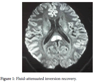
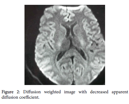
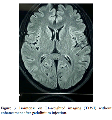
A 27 years old man, with no previous particular medical history was received in our department with seven days psycho-behavioral instability. He presented with incoherent conversations, mystical delusion, spatial disorientation associated to fever (38°C) and cold-like symptoms during this episode. The neurological examination did not show either sensorimotor deficit or meningeal signs. Encephalic Magnetic Resonance Imaging (MRI) has revealed abnormality in the SCC: hyperintense on T2-weighted images (T2WI), fluid-attenuated inversion recovery images (FLAIR) (Figure 1) and diffusion weighted images (Figure 2) with decreased apparent diffusion coefficient (ADC) values; isointense on T1-weighted imaging (T1WI) without enhancement after gadolinium injection (Figure 3).
EEG did not show any paroxysmal activity. Cerebrovascular examination was unremarkable. Immunologic studies for autoimmune disorders were negative. Serum vitamin’s and serum electrolytes levels were normal. At the lumbar puncture: 27 white blood cell (WBC), proteinorachia 0.2 g/l, Glycorachia 4 mmol/l. CRP was strongly positive. Infectious serology was negative (Lyme, brucellosis, HIV, hepatitis B and C, syphilis, Herpes Simplex Virus, adenovirus, coxsackie, cytomegalovirus, echovirus, Influenza A virus, mycoplasma pneumonia) in cerebro-spinal fluid (CSF).
Given the respiratory signs, chest x-ray was realized and did not show any anomaly; analysis of nasal fluid was also made and was positive for Respiratory Syncitial Virus (RSV).
Unfortunately, the search for the RSV genome in the CSF could not be made due to technical problems.
Patient was treated with acyclovir 700 mg intravenous 3 times a day during 14 days. The progress was marked with clinical improvements noticeable from day five.
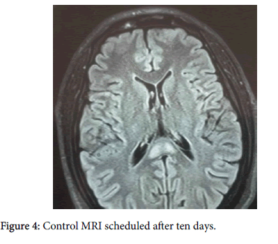
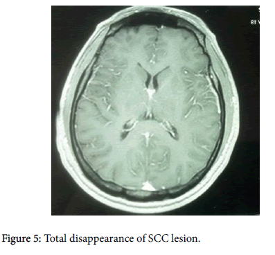
Control MRI scheduled after ten days, has shown negative anomalies notably the almost total disappearance of SCC lesion (Figures 4 and 5).
Discussion
MERS can be the result of many etiologies mainly viral and bacterial infections but also noninfectious etiology such as redundant seizures, anti-epileptic toxicity or brutal breakdown, metabolic disorders (Hypoglycemia-hyponatremia), highly altitude cerebral edema, Kawasaki or Marchiafava-Bignami disease [1].
MERS was described for the first time by Tada et al. [2] Most incriminated germs are as follows: Herpes Simplex Virus, Influenza A virus, mycoplasma pneumonia Adenovirus, Coxsackie, Echoviruses and cytomegalovirus [3].
RSV-a common respiratory virus that usually causes infections of the lungs and respiratory tract; is involved in our observation. We concluded that RSV is necessarily incriminated because of the negativity of all other serologies in CSF, the concomitant positivity of RSV in nasal fluid and the presence of cold signs during the same episode.
Former background knowledge has led us to attribute the first case to this virus.
The exact physiopathology of this entity remains unclear, especially the lesions’ predilection at the level of the SCC. Many physiopathology mechanisms have been suggested: specific affinity of the viral antigen or of the produced antibodies with the receptors of the splenium axons, intramyelinic edema, blood-brain barrier rupture, reversible demyelination or release of arginine vasopressin.
Hyponatremia has been considered by some authors as a major factor of MERS. In Fact, hyponatremia generates decreasing in intercellular osmotic pressure and therefore a transient edema. [4] In his study conducted on 30 patients, published in 2009, Takanashi has found an average low serum level of 131.5 mEq/l [4]. By reviewing working papers as well as the case reported here within, it appears that hyponatremia occurs in less than half of the cases [5,6]. So, hyponatremia is a contributing and precipitating factor but not essential to the development of SCC lesions.
An immune mediated mechanism would be associated with MERS, this is evidenced by the elevation of interleukin-6 in the CSF and antiglutamate 2 receptors in some patients with MERS caused by viral or bacterial infection [7].
A genetic theory has also been suggested because most patients with MERS are of Asian origin [8]. This is the second etiology of acute encephalitis in Japanese children [9]. Imamura and al reported the case of a sibling with MERS reinforcing the genetic hypothesis [10]. Clinical presentation of MERS is nonspecific, without evidence of callosal disconnection syndromes. The most frequently reported clinical signs are: delirious behavior (54%); alteration of consciousness (35%) and epilepsy (33%) [11]. The particularity of this entity resides in the reversible clinical and radiological findings in the periods varying from 2 weeks to one month according to the authors [12]. MRI is essential to perform the diagnosis. It typically shows transient splenial lesions with high-signal-intensity on T2WI and FLAIR, hypo-isointense signal on T1WI sequences without contrast enhancement and restricted diffusion with decreased ADC values indicated from cytotoxic edema [5].
MERS has been subdivided into two sub-entities: Type 1: Lesion limited to the SCC, Type 2: Lesions extended to the whole of corpus callosum or symmetrically extended to the adjacent white matter or extended to both.
Type 1 is more frequent than Type 2, concordant to the radiologic inputs relative to our patient.
Radiologic distinction between these two types reveals a clinical and prognostic interest according to some authors. Thus, Type 2 will be associated to a more severe clinical presentation and a worse prognosis.
MERS must be distinguished from acute disseminated encephalomyelitis (ADEM), posterior reversible encephalopathy syndrome, ischemic stroke or primary CNS lymphoma. Clinical symptomology of ADEM could be similar to MERS but MRI straighten the diagnosis by showing bilateral, asymmetrical and multifocal lesions T2 and FLAIR involving white matter, cerebral cortex, grey matter, and spinal cord with contrast enhancement. The corpus callosum is less affected in ADEM than in MERS. Although, methyl-prednisolone pulse therapy and immunoglobulin were used by many authors; [5] there is still no evidence of efficacy of this treatments on MERS. In the study made by Jing Jing Pan which included 27 patients, only 3 patients were treated with methyl prednisolone pulse therapy, however, all the 27 MERS patients clinically recovered completely [5]. Until now, there is no treatment guideline for MERS.
The prognosis of this entity is favorable. A regression in symptoms has been registered in the majority of cases, which coincide with our observation. Nevertheless, sequels have been reported by Shuo Zhang and et al. and Wen-Xiong Chen et al. in their respective series [1,3].
Conclusion
MERS is often described in children, unlike in adults which is rare. Its etiologies are variable, dominated by infectious causes. Our observation describes the first case of MERS associated to RSV. It allows us to broaden our field of reflexion as regards the infectious etiology in the context of MERS.
References
- Zhang S, Ma Y, Feng J (2015) Clinico radiological spectrum of reversible spleniall esion syndrome (RESLES) in adults: A retrospective study of a rare entity. Medicine (Baltimore) 94: e512.
- Tada H, Takanashi J, Barkovich AJ, Oba H, Maeda M, et al. (2004) Clinically mild encephalitis/encephalopathy with a reversible splenial lesion. Neurology 63: 1854-1858.
- Chen WX, Liu HS, Yang SD, Zeng SH, Gao YY, et al. (2016) Reversible splenial lesion syndrome in children: Retrospective study and summary of case series. Brain Dev 38: 915-927.
- Takanashi J, Tada H, Maeda M, Suzuki M, Terada H (2009) Encephalopathy with a reversible splenial lesion is associated with hyponatremia. Brain Dev 31: 217-220.
- Pan JJ, Zhao YY, Lu C, Hu YH, Yang Y (2015) Mild encephalitis/encephalopathy with a reversible splenial lesion: Five cases and a literature review. NeurolSci 36: 2043-2051.
- Ka A, Britton P, Troedson C, Webster R, Procopis P, et al. (2015) Mild encephalopathy with reversible splenial lesion: An important differential of encephalitis. Eur J Paediatr Neurol 19: 377-382.
- Mori T, Morii M, Kuroiwa Y, Hotsubo T, Fuse S, et al. (2011) Rotavirus encephalitis and cerebellitis with reversible magnetic resonance signal changes. Pediatr Int 53: 252-255.
- Yuan ZF, Shen J, Mao SS, Yu YL, Xu L, et al. (2006) Clinically mild encephalitis/encephalopathy with a reversible splenial lesion associated with Mycoplasma pneumoniae infection. BMC Infect Dis 16: 230.
- Hoshino A, Saitoh M, Oka A, Okumura A, Kubota M, et al. (2012) Epidemiology of acute encephalopathy in Japan, with emphasis on the association of viruses and syndromes. Brain Dev 34: 337-343.
- Imamura T, Takanashi J, Yasugi J, Terada H, Nishimura A (2010) Sisters with clinically mild encephalopathy with a reversible splenial lesion (MERS)-like features; familial MERS? J NeurolSci 290: 153-156.
- Hara M, Mizuochi T, Kawano G, Koike T, Shibuya I, et al. (2011) A case of clinically mild encephalitis with a reversible splenial lesion (MERS) after mumps vaccination. Brain Dev 33: 842-844.
- Takanashi J (2009) Two newly proposed infectious encephalitis/ encephalopathy syndromes. Brain Dev 31: 521-528.
Relevant Topics
- Bacteria Induced Neuropathies
- Blood-brain barrier
- Brain Infection
- Cerebral Spinal Fluid
- Encephalitis
- Fungal Infection
- Infectious Disease in Children
- Neuro-HIV and Bacterial Infection
- Neuro-Infections Induced Autoimmune Disorders
- Neurocystercercosis
- Neurocysticercosis
- Neuroepidemiology
- Neuroinfectious Agents
- Neuroinflammation
- Neurosyphilis
- Neurotropic viruses
- Neurovirology
- Rare Infectious Disease
- Toxoplasmosis
- Viral Infection
Recommended Journals
Article Tools
Article Usage
- Total views: 2941
- [From(publication date):
June-2017 - Aug 30, 2025] - Breakdown by view type
- HTML page views : 2085
- PDF downloads : 856

