Case Report Open Access
Post Malarial Neurological Syndrome: An Uncommon Cause
| Saumya Mittal*, Misri ZK, Rakshith KC and Salony Mittal | |
| Apollo Hospital, Sarita Vihar, Delhi, Uttar Pradesh, India | |
| Corresponding Author : | Saumya Mittal Apollo Hospital, Sarita Vihar Delhi, Uttar Pradesh, India Tel: +919663414840 E-mail: saumyamittal1@gmail.com |
| Received May 14, 2015; Accepted June 15, 2015; Published June 18, 2015 | |
| Citation: Mittal S, Misri ZK, Rakshith KC, Mittal S (2015) Post Malarial Neurological Syndrome: An Uncommon Cause. J Neuroinfect Dis S1:004. doi:10.4172/2314-7326. S1-004 | |
| Copyright: © 2015 Mittal S, et al. This is an open-access article distributed under the terms of the Creative Commons Attribution License, which permits unrestricted use, distribution, and reproduction in any medium, provided the original author and source are credited. | |
| Related article at Pubmed, Scholar Google | |
Visit for more related articles at Journal of Neuroinfectious Diseases
Abstract
Post Malarial Neurological Syndrome a manifestation of cerebral malaria is known to be caused by plasmodium falciparum. It’s characterized by aphasia, tremors, myoclonus, psychiatric manifestations and white matter patchy changes in MRI. Post malarial neurological syndrome causes mild neurological deficits to severe encephalopathies. We present a case with this syndrome and all its neuroradiological and EEG manifestations caused by plasmodium vivax. Vivax malaria is known to have a more benign course. In our review, we found no reports suggestive of post malarial neurological syndrome caused by plasmodium vivax. We therefore report the case and review the manifestations of Post Malarial Neurological Syndrome.
| Keywords |
| Plasmodium vivax; Post malarial neurological syndrome; Cerebral malaria |
| Introduction |
| Malaria, a major health problem in India, causes approximately 300-500 million cases and 1.2-7 million deaths. Approximately 40% of cases of malaria, outside of Africa, occur in India. And plasmodium vivax causes 60-70% of these cases. Cerebral malaria, a dreaded complication of malaria, is commonly caused by plasmodium falciparum [1]. |
| Severe malaria is usually caused by plasmodium falciparum. It is characterized by cerebral malaria, severe normocytic anemia, renal failure, pulmonary edema, hypoglycemia, circulatory collapse, spontaneous bleeding, DIC, repeated seizures and malarial hemoglobinuria [2]. |
| Case |
| The patient, a 60 year old hypertensive and alcoholic gentleman, presented to the hospital with sudden onset of loss of consciousness and first episode of convulsions. He presented to the hospital in altered sensorium, GCS-3, with hypotension (BP- 90/60 mm Hg) and tachycardia (HR- 128/min). No neck stiffness was noted. The patient, treated with antiepileptics and ionotropes was intubated and ventilated. |
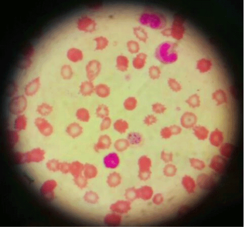
| He was found to have thrombocytopenia (27000/cmm), azotemia (urea- 60, creatinine- 2.3), and raised liver enzymes (AST- 373, ALT- 258, ALP- 65). Peripheral smear isolated schizonts and gametocytes of plasmodium vivax (Figure 1). |
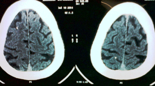
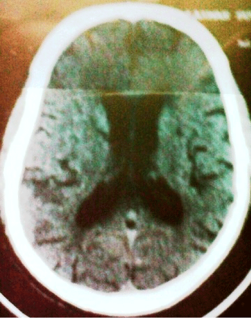
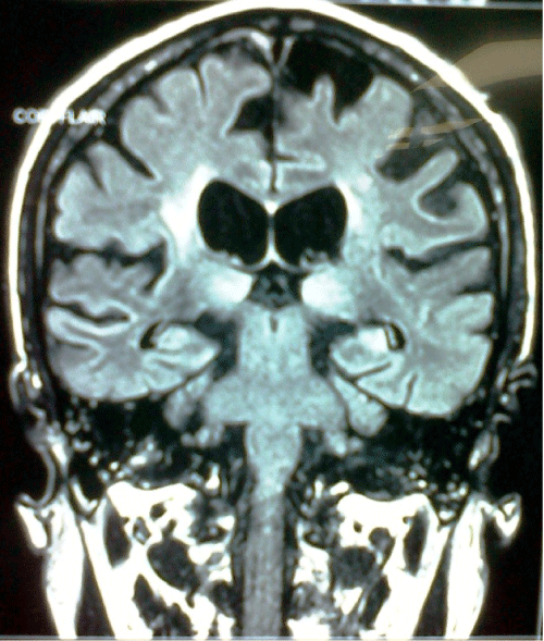
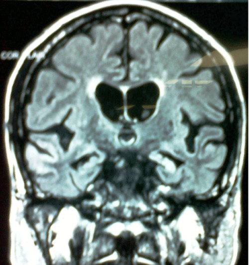
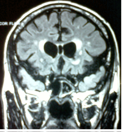
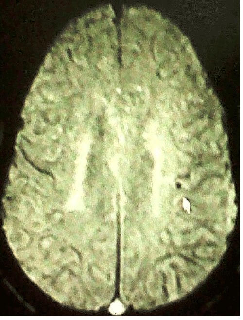
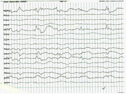
| CAT scan of the brain showed Calcified lesions seen in temporoparietal region (Figure 2) and periventricular white matter hyperintensities (Figure 3). MRI was suggestive of B/L thalamus involvement, ventricular dilatation (Figure 4), temporal and hippocampal hyperintensities (Figure 5), anterior temporal involvement (Figure 6), and micro hemorrhages (Figure 7). The EEG showed slowing of waves (Figure 8). |
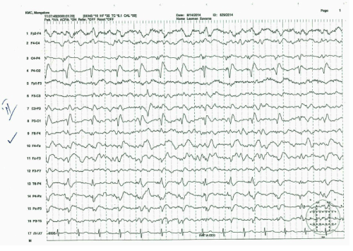
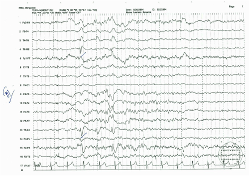
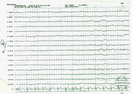
| The patient diagnosed with cerebral malaria and initiated on treatment. The patient developed first episode of fever with chills and rigors 12 hours after admission to the hospital. He apparently improved, but developed recurrent seizures (generalized and focal) after a week. The EEG was suggestive of a spike wave pattern (Figure 9) that became less frequent after 4 days (Figure 10). Over a period of time, sensorium and motor power improved but continued to have mild cognitive dysfunction, tremors, mild aphasia and inappropriate response to command. On review in the opd, the patient was neurologically stable and the EEG was normal (Figure 11). |
| Discussion |
| Cerebral malaria is a serious acute febrile diffuse bihemispheric encephalopathy in which localizing signs are absent or transitory and plasmodium falciparum is demonstrable. It manifests with acute widespread bilateral, symmetrical encephalopathy producing seizures that may accompany or precede loss of consciousness (focal or generalized), hypertonia, brisk deep tendon reflexes, extensor plantar response, extensor posturing, decerebrate rigidity/decorticate rigidity, coma (with GCS<9) and sustained upward deviation of eyes [2]. Prognosis usually worsens as the duration of coma increases and the patient may have long term cognitive impairment [3]. |
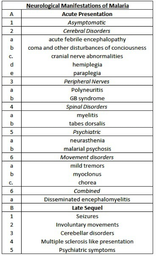
| Neurological manifestations can be divided into acute and late sequel (Figure 12). Sequels are commoner in children and include extrapyramidal rigidity, tremors, cerebellar ataxia, brief reactive psychosis, hemiplegia, cortical blindness and cranial nerve abnormalities [3]. |
| Post malarial neurological syndrome is characterized by aphasia, tremors, myoclonus, psychiatric manifestations (all of which are responsive to steroids) and white matter patchy changes in MRI. Post malarial neurological syndrome causes mild neurological deficits to severe encephalopathies. It may be classified into three subtypes: 1. Mild or localized form characterized by isolated cerebellar ataxia or postural tremor [2]. Diffuse, moderate encephalopathy with acute confusional or epileptic states [3]. Severe corticosteroid responsive encephalopathy causing confusion, focal deficits, tremors, cerebellar ataxia, myoclonus [2,4]. |
| The possible mechanism causing this may be cerebral hypoxemia due to obstruction of cerebral microvasculature by parasitized erythrocytes. This may lead to presence of petechial hemorrhages. This sequestration seems to be essential to the pathogenesis. Alternatively, immune mechanisms may be involved. CSF and plasma concentrations of cytokines may be raised e.g. TNF-α, IL-2 and IL-6. The levels of the cytokines may be persistently raised despite eradication of the parasites. Another possible cause is the antibody induced damage due to molecular mimicry between antigens of the plasmodium falciparum and the antigens in nervous system [2,5]. |
| Cerebrospinal fluid findings are variable and show raised pressure, cell count and proteins. Rarely, xanthochromia may be seen. CAT scan may be normal. Alternatively, diffuse cerebral edema may be seen. Bilateral symmetric non enhancing thalamic and/or cerebellar hypodensities or bilateral thalamic hemorrhagic infarcts can occur. Widespread low density areas suggestive of ischemic damage and consistent with a critical reduction in cerebral perfusion pressure are frequently seen. MRI may be suggestive of hemorrhagic cortical lesions thought to be infarct. Cerebellar and thalamic lesions are better appreciated. Diffuse or watershed zone (ACA-MCA, MCA-PCA) ischemic lesions may be seen. Diffuse white matter edema or gliosis may be seen. This may regress with antimalarial treatment. Central pontine myelinosis or myelinosis of upper medulla may be seen [2,6]. |
| EEG may be suggestive of a poor prognosis if it shows a slow frequency, has a background asymmetry, and if it depicts burst suppression or interictal discharges [2]. |
| Conclusion |
| Vivax malaria is known to have a more benign course. Plasmodium vivax malaria is known to run a benign course. However, a change in the genes, or a change in the vector, or the drug resistance may be giving rise to parasites that are leading to a change in the clinical profile. It was initially suspected that such cases may be due to coinfection of plasmodium vivax with plasmodium falciparum. But, using the newer malarial antigen test and nucleic acid amplification technique, this theory has been disproved [5]. |
| In our review, we found case reports suggestive of cerebral malaria caused by plasmodium vivax. However, we found no reports suggestive of post malarial neurological syndrome caused by plasmodium vivax. All reports we came across were caused by plasmodium falciparum. We present a case where the syndrome is caused by plasmodium vivax, when it is usually caused by plasmodium falciparum. |
| Acknoledgement |
| Acknowledgement to Dr Chakrapani, Professor Medicine and Associate Dean, KMC Mangalore for his guidance. |
References
- Neki NS (2013) Cerebral malaria caused by Plasmodium vivax. JIACM 14: 69-70.
- WHO (2000) Severe Falciparum Malaria. Transitions of the Royal Society of Tropical Medicine and Hygeine.
- Idro R, Jenkins NE, RJC Newton C (2005) Pathogenesis, clinical features, and neurological outcome of cerebral malaria. The Lancet Neurology 4: 827-840.
- Marsala SZ, Ferracci F, Cecotti L (2006) Post-malarial neurological syndrome: clinical and laboratory findings in one patient. NeurolSci27: 442-4.
- Limaye CS, Londhey VA, Nabar ST (2012)The Study of Complications of Vivax Malaria in Comparison with Falciparum Malaria in Mumbai. JAPI 60: 15-8.
- Potchena MJ, Birbeckb GL, DeMarcoa K(2010) Neuroimaging findings in children with retinopathy-confirmed cerebral malaria. European Journal of Radiology 74: 262-8.
Figures at a glance
 |
 |
 |
 |
| Figure 1 | Figure 2 | Figure 3 | Figure 4 |
 |
 |
 |
 |
| Figure 5 | Figure 6 | Figure 7 | Figure 8 |
 |
 |
 |
 |
| Figure 9 | Figure 10 | Figure 11 | Figure 12 |
Relevant Topics
- Bacteria Induced Neuropathies
- Blood-brain barrier
- Brain Infection
- Cerebral Spinal Fluid
- Encephalitis
- Fungal Infection
- Infectious Disease in Children
- Neuro-HIV and Bacterial Infection
- Neuro-Infections Induced Autoimmune Disorders
- Neurocystercercosis
- Neurocysticercosis
- Neuroepidemiology
- Neuroinfectious Agents
- Neuroinflammation
- Neurosyphilis
- Neurotropic viruses
- Neurovirology
- Rare Infectious Disease
- Toxoplasmosis
- Viral Infection
Recommended Journals
Article Tools
Article Usage
- Total views: 15228
- [From(publication date):
specialissue-2015 - Aug 24, 2025] - Breakdown by view type
- HTML page views : 10469
- PDF downloads : 4759
