 Spanish
  Chinese
  Russian
  German
  French
  Japanese
  Portuguese
  Hindi
Hindi Our Group organises 3000+ Global Conferenceseries Events every year across USA, Europe & Asia with support from 1000 more scientific Societies and Publishes 700+ Open Access Journals which contains over 50000 eminent personalities, reputed scientists as editorial board members.
Open Access Journals gaining more Readers and Citations
700 Journals and 15,000,000 Readers Each Journal is getting 25,000+ Readers
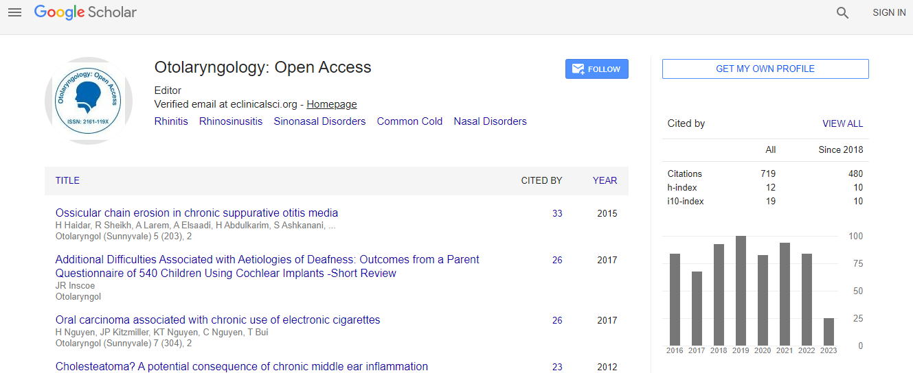
Google Scholar citation report
Citations : 717
Otolaryngology: Open Access received 717 citations as per Google Scholar report
Otolaryngology: Open Access peer review process verified at publons

Indexed In
- Index Copernicus
- Google Scholar
- Sherpa Romeo
- Open J Gate
- Genamics JournalSeek
- RefSeek
- Hamdard University
- EBSCO A-Z
- OCLC- WorldCat
- Publons
- Geneva Foundation for Medical Education and Research
- ICMJE
Useful Links
Recommended Journals
Related Subjects
Share This Page
The importance and timing of optic canal exploration and decompression during endoscopic endonasal resection of tuberculum sella and planum sphenoidale meningiomas
Global Summit and Medicare Expo on Head & Neck Surgery
Attia M1, 2, Kandasamy J1, Jakimovski D1, Bedrosian J1, Alimi M1, Lee D L1, Anan V K1 and Schwartz T H1
1Weill Cornell Medical College, USA 2Rambam Health Care Campus, Israel
ScientificTracks Abstracts: Otolaryngol (Sunnyvale)
Abstract
Background: Suprasellar meningiomas often invade the optic canals (OCs). The feasibility of removing these tumors through a minimal-access endonasal route has been demonstrated but the importance, safety and timing of OC exploration and decompression are not well described. Objective: To create a simple decision-tree algorithm for OC exploration and decompression in the endonasal endoscopic surgery for planum sphenoidale and tuberculum sella meningiomas. Methods: We identified a consecutive series of 8 planum sphenoidale and tuberculum sella meningiomas resected endonasally. ��?Late� OC exploration and decompression was performed in 4 of 8 patients. The extent of resection, visual outcome and complications were recorded. Results: Five patients had OC invasion on magnetic resonance imaging. Endoscopic inspection did not reveal additional OC invasion. The OC was opened bilaterally in 2 patients and unilaterally in 2 patients. Gross total resection was achieved in 6 of 7 patients in whom it was the goal. Vision improved in 3 patients (3 of 3 OCs opened) and was stable in 4 (1 of 4 OCs opened). In 1 patient, the bitemporal hemianopsia improved but there was unilateral deterioration (no OC invasion) because the tumor was extremely adherent to 1 optic nerve. After an average follow-up of 20.9 months, all patients had Glasgow Outcome Scale score of 5 and there were no cerebrospinal fluid leaks. Conclusion: Exploration and decompression of the OC are feasible, safe and important to optimize visual outcome and to minimize recurrence in planum sphenoidale and tuberculum sella meningiomas resected endonasally. It may not be important to open the canal early during surgery because tumor debulking can be performed without manipulating the optic nerves. Early decompression however is technically feasible.Biography
Attia M is a Board Certified Neurosurgeon in the Department of Neurosurgery at Rambam Medical Center in Haifa, Israel since 2012. His specialty focuses on skull base and cerebrovascular surgery. He is serving as Residency Program Director and as attending Neurosurgeon in his Department. He has graduated from the Hebrew University and Hadassah Medical School in 2001 and completed Residency in Neurosurgery at Chaim Sheba Medical Center in Tel HaShomer in 2007. Afterwards, he has accomplished a three years Clinical Fellowship specializing in Skull Base and Cerebrovascular Neurosurgery at the University of Washington and University of Illinois at Chicago and at Hadassah Medical Center. He has then served as an Attending Neurosurgeon at Hadassah.
Email: moshe.attia24@gmail.com