Research Article Open Access
Lumbar Facet Joint Injections: Is CT Guided Intra-Articular Needle Position Advantageous?
| Lilach Goldstein1, Karin Ariche2, Elon Eisenberg2,3*, Roi Treister4,3 and Rimma Geller2,5 | |
| 1Department of Neurology, Rabin Medical Center, Campus Beilinson, Petach Tikva, Israel | |
| 2Institute of Pain Medicine, Rambam Health Care Campus, Haifa, Israel | |
| 3The Technion, Israel Institute of Technology, Haifa, Israel | |
| 4Nerve Injury Unit, Department of Neurology, Massachusetts General Hospital & Harvard Medical School, Boston, USA | |
| 5Department of Anesthesiology, Rambam Health Care Campus, Haifa, Israel | |
| Corresponding Author : | Elon Eisenberg Institute of Pain Medicine Rambam Health Care Campus, Haifa Israel Haifa 31096, Israel Tel: +972-4-7772234 Fax: +972-4-7773505 E-mail: e_eisenberg@rambam.health.gov.il |
| Received December 20, 2014; Accepted March 16, 2015; Published March 19, 2015 | |
| Citation: Goldstein L, Ariche K, Eisenberg E, Roi Treister R, Geller R (2015) Lumbar Facet Joint Injections: Is CT Guided Intra-Articular Needle Position Advantageous?. J Pain Relief 4:178. doi: 10.4172/2167-0846.1000178 | |
| Copyright: © 2015 Goldstein L, et al. This is an open-access article distributed under the terms of the Creative Commons Attribution License, which permits unrestricted use, distribution, and reproduction in any medium, provided the original author and source are credited. | |
Visit for more related articles at Journal of Pain & Relief
Abstract
Background: Although facet joint injections of corticosteroids and local anesthetics are commonly performed for treating low back pain (LBP), their effectiveness remains questionable. This is partially due to lack of consensus regarding the correct needle-tip location within or nearby the facet joint. Purpose: The present study was designed to test if computerized tomography (CT) guided intra-articular needle position yields better results than peri-articular position of the needle, while performing lumbar facet joints injections for chronic LBP. Study Design/Setting: A prospective, randomized controlled trial conducted in A university hospital based pain clinic Patient sample: Forty-nine patients with chronic LBP related to facet joint arthropathy. Outcome measures: Scales of pain severity, analgesic drug consumption, lumbar motion, disability and patient's global impression of improvement. Methods: Patients were randomized to receive CT-guided intraarticular (n= 26) or periarticular (n=23) needle-tip positions during facet joint injections steroids and local anesthetics. Selection of the facet joint for injection was based on medical history, physical findings, CT scan, and bone scintigraphy. Patients were followed for eight weeks. Results: Although all outcome measures improved significantly from baseline throughout the entire follow-up period, none of them differed statistically or clinically between the two study groups. Conclusions: Facet joint injections of corticosteroids and local anesthetics provide short-term improvements in pain and disability in patients with chronic low back pain due to facet joints arthropathy. However, efforts to precisely locate the needle-tip within the facet joint – as oppose to perform a peri-articular injection - are not advantageous.
Purpose: The present study was designed to test if computerized tomography (CT) guided intra-articular needle position yields better results than peri-articular position of the needle, while performing lumbar facet joints injections for chronic LBP.
Study Design/Setting: A prospective, randomized controlled trial conducted in A university hospital based pain clinic
Patient sample: Forty-nine patients with chronic LBP related to facet joint arthropathy.
Outcome measures: Scales of pain severity, analgesic drug consumption, lumbar motion, disability and patient's global impression of improvement.
Methods: Patients were randomized to receive CT-guided intraarticular (n= 26) or periarticular (n=23) needle-tip positions during facet joint injections steroids and local anesthetics. Selection of the facet joint for injection was based on medical history, physical findings, CT scan, and bone scintigraphy. Patients were followed for eight weeks.
Results: Although all outcome measures improved significantly from baseline throughout the entire follow-up period, none of them differed statistically or clinically between the two study groups.
Conclusions: Facet joint injections of corticosteroids and local anesthetics provide short-term improvements in pain and disability in patients with chronic low back pain due to facet joints arthropathy. However, efforts to precisely locate the needle-tip within the facet joint – as oppose to perform a peri-articular injection - are not advantageous.
Facet interventions represent the second most common type of procedure performed in pain management centers throughout the United States [1]. These interventions include, among others, facet joint injections of corticosteroids that are aimed to reduce synovial inflammation and pain [1]. Although several studies have assessed the analgesic effect of facet joint injections, their efficacy remains uncertain [2-6]. Staal et al. [2] conducted a systematic review on the effectiveness of injection therapy for subacute and chronic low back pain. They concluded that there is conflicting evidence as to whether facet joint injections with corticosteroids are more effective than placebo injections for pain reduction and improvement of disability [2]. Falco et al. [3] also concluded in their recent review that the evidence for the efficacy of IA injections is limited.
The mixed results regarding the efficacy of facet joint injections may be a consequence of inconsistent parameters used in patient selection for the procedure. For example, it has been shown that positive bone scans (with SPECT) helped in identifying patients who would benefit from a facet joint injection, thus leading to better clinical outcomes [7-9]. In addition, differences in injection techniques, including the volume and type of injected drugs and guidance techniques used, can also contribute to the outcome. Another important factor that may account for the inconsistency is the localization of the needle, namely whether it is placed inside the joint (intra-articular; IA) or nearby the joint (peri-articular; PA). Lilius et al. [4] compared the effectiveness of fluoroscopy-guided (IA) versus PA injections of steroids and local anesthetics. Their results showed no differences between these two injection techniques. On the other hand, Lynch et al. [10] demonstrated that fluoroscopy-guided injections into the joints were far more effective than extra-articular injections for achieving long-term pain relief.
In an effort to further address this issue, the present study was aimed to test if computerized tomography (CT) guided intra-articular needle position yields better results than peri-articular position of the needle, while performing lumbar facet joints injections of corticosteroid and local anesthetics for chronic facet arthropathy-induced LBP.
Injections with IA needle positions were performed in 26 patients and with PA needle positions in 23 patients. No differences between the two study groups were found in any of the demographic characteristics, including gender, working status, age, or education. In 32 patients, the injections were performed bilaterally, for a total of 64 injected joints, while each of the other 17 patients received only one injection. No differences between the treatment groups in the number of patients who received bilateral injections were found (IA: 18 patients; PA: 14 patients; p=0.625). A total of 81 joints were injected, with 44 IA needle positions and 37 PA needle positions. A description of the subjects' characteristics in the two study groups is presented in Table 1.
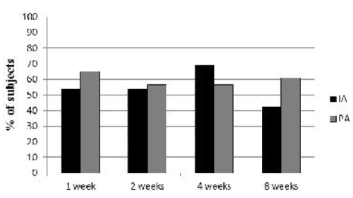
Chi-Square tests failed to demonstrate significant differences between the two treatment groups at any of the follow-up time points in the percentages of patients who decreased their analgesic drug consumption from baseline (week 1: p=0.419; week 2: p=0.851; week 4: p=0.357; week 8: p=0.195).
From the mechanism-related viewpoint, the fact that no differences were found between IA and PA needle positions in the current study may imply that underlying mechanisms additional to joint inflammation are involved in facet pain generation. In line with this assumption, McCall et al. [13] found no difference in the pain referral areas induced in healthy subjects by IA and PA injections of hypertonic saline.
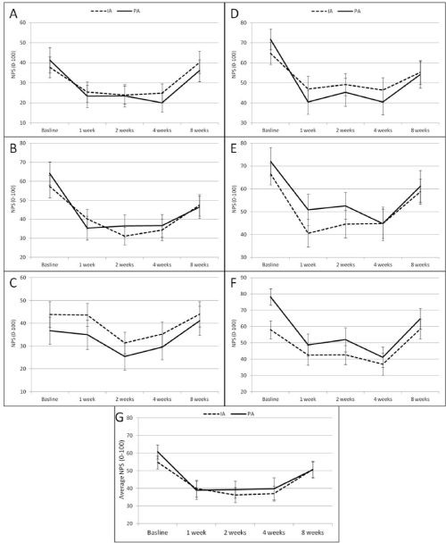
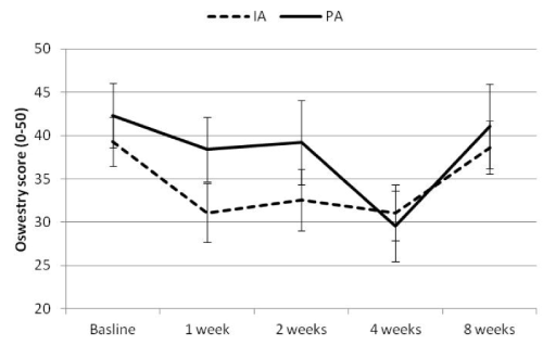
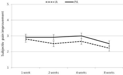
Regardless of the similarities between the effects of the IA and the PA needle positions, an additional finding of the current study relates to the duration of the analgesic effect of these facet injections. Strong pain relief was evident in all outcome measures for up to four weeks after the injections. This effect subsequently subsided, and at the eight-week follow-up, only the primary outcome measure (overall pain intensity) remained significantly different from the baseline measurement. These results are similar to those reported by Chaturvedi et al. [14]. In their trial, most patients reported significant pain relief immediately after the procedure. The number of patients increased slightly at one week and reached a peak at four weeks, by which time as many as 93.3% of patients had responded. Pain relief declined at 12 and 24 weeks [14]. Marks et al. [15] found a similar short-lived response [15]. In contrast, a few other studies have reported longer analgesic effects, lasting for three months or more [10,16-18]. Differences in the duration of effect can be explained by differences in the drug type, volume, and dosage used in the different studies. In addition, variations in study methodology may also contribute to these inconsistencies. For example, in our study, questionnaires were completed four times during the eight weeks of follow-up, whereas in Fotiadou et al.'s [16] study, in which longer analgesic effects were demonstrated, questionnaires were completed only once at three months following treatment.
Both Carette et al. [19] and Lilius et al. [4] found no difference in pain reduction between steroid and placebo injections. Yet, in both studies, the placebo arm included an IA injection of normal saline. Notably, saline injection into the facet space may have therapeutic properties, given that inflammatory mediators can presumably be expelled following the injection. Indeed, normal saline has been shown to provide better pain relief than expected with a true placebo in a multitude of invasive procedures [1,3].
Determination of diagnosis and selection of the joint for injection are important factors in the treatment outcome. Some authors claim that a positive response to medial branch block is the only accurate test for the diagnosis of faced pain [20]. However, others find clinical and radiological findings as sufficient for establishing the diagnosis correctly [7-9,18,21,22]. In our study we have opted to use the second set of criteria and the diagnosis of facet pathology was based on strict clinical and radiological parameters, including medical history and physical examination, as well as CT and bone scans.
Two study limitations should be considered. First, since no placebo control group was included in the present study, one cannot rule out the possibility that the improvement in the outcome measures found may be attributable to a placebo effect. A future study with random IA versus PA needle tip locations should include injections of an active drug or a placebo for excluding this possibility. Second, some joints were injected with half of the quantity of drugs as compared to others, due to the fact that in some patients only one joint was injected and in others two joints received the treatment. This may have reduced the consistency of the results. However, since this was done in similar numbers of patients from both groups, it is unlikely to have skewed the comparisons between the outcomes of the two injection techniques.
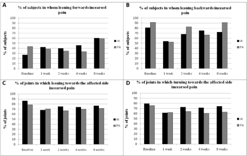
Lastly, two additional points deserve consideration. First, trends for differences between the groups in baseline pain intensities in five of the six tested positions (all but sitting) was found, with higher values for the IA group. Similarly, the magnitudes of drop in pain intensities are seemingly larger for the IA as compared to the PA group, in four of these positions. Although these trends have not reached statistical significance, they seem to justify larger-sized future trials to assure that no real differences in the efficacy between these interventions indeed exist. Second, a large drop in pain intensity in relation to those five positions occurred during the first post-injection week, whereas not similar drop was seen for the sitting position. This can be partially explained by the low baseline pain intensity during sitting relative to most other positions, which is typical for patients with facet pain.
In summary, the current study revealed no differences between IA and PA needle positions during facet joint injections. PA needle position is an easier technique that usually necessitates less radiation for verification of the needle position. Moreover, our clinical experience has demonstrated that in some cases, there are difficulties in inserting the needle into the joint space due to anatomical changes. Since both methods result in similar outcomes, the use of PA needle positions should be considered.
References
- Cohen SP, Raja SN (2007) Pathogenesis, diagnosis, and treatment of lumbar zygapophysial (facet) joint pain. Anesthesiology106: 591-614.
- Staal JB, de Bie RA, de Vet HC, Hildebrandt J, Nelemans P (2009) Injection therapy for subacute and chronic low back pain: an updated Cochrane review. Spine (Phila Pa 1976) 34: 49-59.
- Falco FJ, Manchikanti L, Datta S, Sehgal N, Geffert S, et al. (2012) An update of the effectiveness of therapeutic lumbar facet joint interventions. Pain Physician 15: E909-953.
- Lilius G, Laasonen EM, Myllynen P, Harilainen A, Grönlund G (1989) Lumbar facet joint syndrome. A randomised clinical trial. J Bone Joint Surg Br 71: 681-684.
- van Kleef M, Vanelderen P, Cohen SP, Lataster A, Van Zundert J, et al. (2010) 12. Pain originating from the lumbar facet joints. Pain Pract 10: 459-469.
- Peh W (2011) Image-guided facet joint injection. Biomed Imaging Interv J 7: e4.
- Holder LE, Machin JL, Asdourian PL, Links JM, Sexton CC (1995) Planar and high-resolution SPECT bone imaging in the diagnosis of facet syndrome. J Nucl Med 36: 37-44.
- Dolan AL, Ryan PJ, Arden NK, Stratton R, Wedley JR, et al. (1996) The value of SPECT scans in identifying back pain likely to benefit from facet joint injection. Br J Rheumatol 35: 1269-1273.
- Pneumaticos SG, Chatziioannou SN, Hipp JA, Moore WH, Esses SI (2006) Low back pain: prediction of short-term outcome of facet joint injection with bone scintigraphy. Radiology 238: 693-698.
- Lynch MC, Taylor JF (1986) Facet joint injection for low back pain. A clinical study. J Bone Joint Surg Br 68: 138-141.
- Fairbank JC, Pynsent PB (2000) The Oswestry Disability Index. Spine (Phila Pa 1976) 25: 2940-2952.
- Davidson M, Keating JL (2002) A comparison of five low back disability questionnaires: reliability and responsiveness. PhysTher 82: 8-24.
- McCall IW, Park WM, O'Brien JP (1979) Induced pain referral from posterior lumbar elements in normal subjects. Spine (Phila Pa 1976) 4: 441-446.
- Chaturvedi A, Chaturvedi S, Sivasankar R (2009) Image-guided lumbar facet joint infiltration in nonradicular low back pain. Indian J Radiol Imaging 19: 29-34.
- Marks RC, Houston T, Thulbourne T (1992) Facet joint injection and facet nerve block: a randomised comparison in 86 patients with chronic low back pain. Pain 49: 325-328.
- Fotiadou A, Wojcik A, Shaju A (2012) Management of low back pain with facet joint injections and nerve root blocks under computed tomography guidance. A prospective study. Skeletal Radiol 41: 1081-1085.
- Schulte TL, Pietilä TA, Heidenreich J, Brock M, Stendel R (2006) Injection therapy of lumbar facet syndrome: a prospective study. ActaNeurochir (Wien) 148: 1165-1172.
- Lakemeier S, Lind M, Schultz W, Fuchs-Winkelmann S, Timmesfeld N, et al. (2013) A comparison of intraarticular lumbar facet joint steroid injections and lumbar facet joint radiofrequency denervation in the treatment of low back pain: a randomized, controlled, double-blind trial. AnesthAnalg 117: 228-235.
- Carette S, Marcoux S, Truchon R, Grondin C, Gagnon J, et al. (1991) A controlled trial of corticosteroid injections into facet joints for chronic low back pain. N Engl J Med 325: 1002-1007.
- Cohen SP, Huang JH, Brummett C (2013) Facet joint pain--advances in patient selection and treatment. Nat Rev Rheumatol 9: 101-116.
- Silbergleit R, Mehta BA, Sanders WP, Talati SJ (2001) Imaging-guided injection techniques with fluoroscopy and CT for spinal pain management. Radiographics 21: 927-939.
- Gorbach C, Schmid MR, Elfering A, Hodler J, Boos N (2006) Therapeutic efficacy of facet joint blocks. AJR Am J Roentgenol 186: 1228-1233.
Tables and Figures at a glance
| Table 1 |
Figures at a glance
 |
 |
 |
 |
 |
| Figure 1 | Figure 2 | Figure 3 | Figure 4 | Figure 5 |
Relevant Topics
- Acupuncture
- Acute Pain
- Analgesics
- Anesthesia
- Arthroscopy
- Chronic Back Pain
- Chronic Pain
- Hypnosis
- Low Back Pain
- Meditation
- Musculoskeletal pain
- Natural Pain Relievers
- Nociceptive Pain
- Opioid
- Orthopedics
- Pain and Mental Health
- Pain killer drugs
- Pain Mechanisms and Pathophysiology
- Pain Medication
- Pain Medicine
- Pain Relief and Traditional Medicine
- Pain Sensation
- Pain Tolerance
- Post-Operative Pain
- Reaction to Pain
Recommended Journals
Article Tools
Article Usage
- Total views: 14407
- [From(publication date):
March-2015 - Aug 20, 2025] - Breakdown by view type
- HTML page views : 9832
- PDF downloads : 4575
