Our Group organises 3000+ Global Conferenceseries Events every year across USA, Europe & Asia with support from 1000 more scientific Societies and Publishes 700+ Open Access Journals which contains over 50000 eminent personalities, reputed scientists as editorial board members.
Open Access Journals gaining more Readers and Citations
700 Journals and 15,000,000 Readers Each Journal is getting 25,000+ Readers
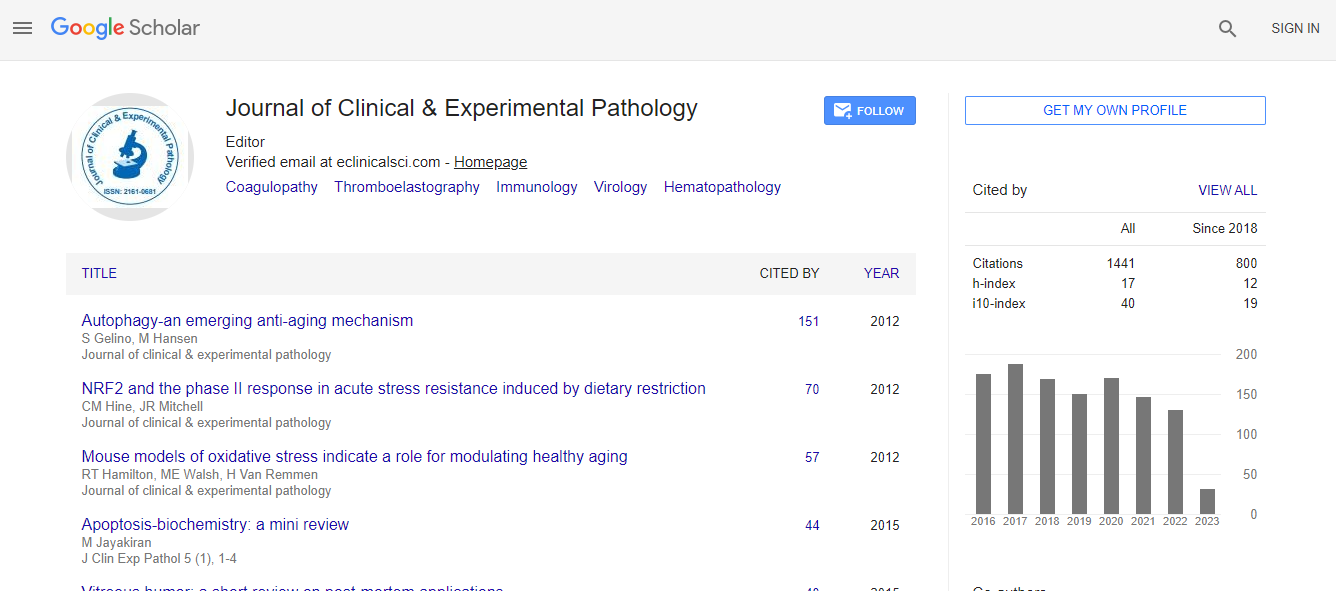
Google Scholar citation report
Citations : 2975
Journal of Clinical & Experimental Pathology received 2975 citations as per Google Scholar report
Journal of Clinical & Experimental Pathology peer review process verified at publons
Indexed In
- Index Copernicus
- Google Scholar
- Sherpa Romeo
- Open J Gate
- Genamics JournalSeek
- JournalTOCs
- Cosmos IF
- Ulrich's Periodicals Directory
- RefSeek
- Directory of Research Journal Indexing (DRJI)
- Hamdard University
- EBSCO A-Z
- OCLC- WorldCat
- Publons
- Geneva Foundation for Medical Education and Research
- Euro Pub
- ICMJE
- world cat
- journal seek genamics
- j-gate
- esji (eurasian scientific journal index)
Useful Links
Recommended Journals
Related Subjects
Share This Page
Data fusion: A new paradigm for 21st century diagnostics
5th International Conference on Pathology
John E Tomaszewski
State University of New York at the University at Buffalo, USA
Keynote: J Clin Exp Pathol
Abstract
Computational advances offer the promise of enabling the quantitative analysis of structural data at all levels of scale. In Pathology high-resolution cellular imaging methods including histology, super resolution optical, and electron microscopic examination can be married with other data using the new analytics of machine vision and machine learning. The computational analysis of structure offers incredible new tools with which to quantitatively mine the data within both macroscopic structure (101) and microscopic (10-6 to -9) worlds and integrate those data with other modes including molecular and cell biology information. In our work we seek to use quantitative histological image analysis for modeling complex biological systems. We do this starting with a fundamental hypothesis which is that a high-resolution image is a self-organizing set of data that uniquely represents all of the genes, all of the molecules, and all of the cells captured at one point in time. In other words, a histological image is what it is for very specific reasons and those reasons are the relationships amongst the genomics, epigenomics, proteomics, metabolomics, and all the ��?omics� that go into making that image. The promise of quantitative histological image analysis lies in the hypothesis that the linkages relating all of the molecular events contributing to an image are still extant and minable. This keynote address will review some of our work in this new domain of data fusion and integrated diagnostics.Biography
John E Tomaszewski received his MD from the University of Pennsylvania, Philadelphia, PA in 1977. He is currently Professor and Chair of Pathology and Anatomical Sciences at the State University of New York at the University at Buffalo. He has published more than 300 papers in the domains of pathology diagnostics and the application of quantitative imaging to diagnostics.
Email: johntoma@buffalo.edu

 Spanish
  Chinese
  Russian
  German
  French
  Japanese
  Portuguese
  Hindi
Hindi 
