Our Group organises 3000+ Global Conferenceseries Events every year across USA, Europe & Asia with support from 1000 more scientific Societies and Publishes 700+ Open Access Journals which contains over 50000 eminent personalities, reputed scientists as editorial board members.
Open Access Journals gaining more Readers and Citations
700 Journals and 15,000,000 Readers Each Journal is getting 25,000+ Readers
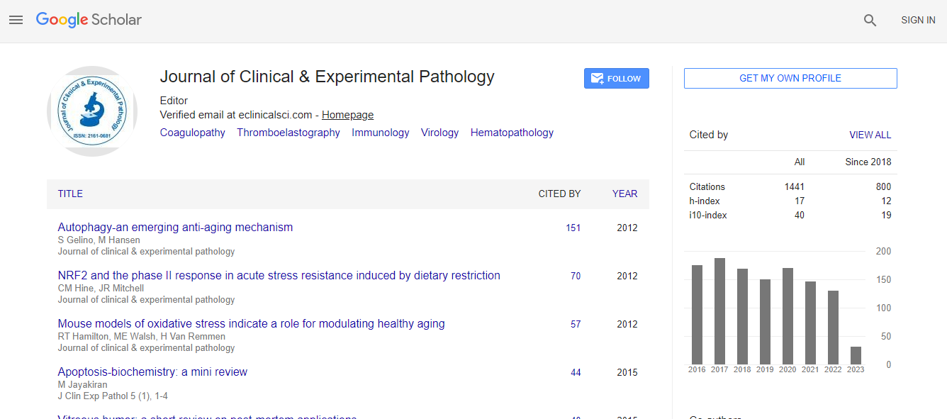
Google Scholar citation report
Citations : 2975
Journal of Clinical & Experimental Pathology received 2975 citations as per Google Scholar report
Journal of Clinical & Experimental Pathology peer review process verified at publons
Indexed In
- Index Copernicus
- Google Scholar
- Sherpa Romeo
- Open J Gate
- Genamics JournalSeek
- JournalTOCs
- Cosmos IF
- Ulrich's Periodicals Directory
- RefSeek
- Directory of Research Journal Indexing (DRJI)
- Hamdard University
- EBSCO A-Z
- OCLC- WorldCat
- Publons
- Geneva Foundation for Medical Education and Research
- Euro Pub
- ICMJE
- world cat
- journal seek genamics
- j-gate
- esji (eurasian scientific journal index)
Useful Links
Recommended Journals
Related Subjects
Share This Page
Gross digital pathology advancement
5th International Conference on Pathology
Izak Dimenstein
Info-Genesis, USA
Posters & Accepted Abstracts: J Clin Exp Pathol
Abstract
The development of digital pathology in the surgical pathology laboratory requires attention not only to the digitalized micro slide. Gross digital pathology can contribute to the pathologist√ʬ?¬?s report at different stages of the diagnostic process, including the gross section close-up images directly corresponding to the micro slide. Although all modern photo stand cameras are able to provide a satisfactory macro image, these close-ups images require a special device. The prototype of a portable device is proposed for close-up images capturing selected gross sections. The device includes a camera mounted on the grossing board. The size and configuration of the board depends on the predominant type of specimens. The camera is focused on certain foreshortened positions of a securely immobilized specimen on the special cutting platform. Exclusive close-up photography has important requirements, such as intensive illumination with minimal shadows and smaller depth of field to display details in focus. The device supposed to work in a hectic assembly line like surgical pathology laboratory environment; therefore the camera√ʬ?¬?s operations including Wi-Fi variants are limited to a minimal number of focused positions. The board should be convenient and easily moved from one grossing station to another. Camera√ʬ?¬?s and lightning√ʬ?¬?s holders with clamps can be handy detached from the board for fast cleanup.Biography
Izak Dimenstein Graduated from Mechnikov Medical Academy in Leningrad, former USSR, in 1964 and completed my PhD program at the same institution in 1969. He has worked as a clinical and anatomical pathologist in Leningrad, USSR. Since 1995, he worked as a pathologists’ assistant and grossing technologist at Mount Sinai Chicago Hospital and Loyola University Chicago Medical Center developed a website entitled “Grossing Technology in Surgical Pathology” (www. grossing-technology.com) in 2002. He has retired from Loyola University Chicago Medical Center in 2008. Currently, his main area of interest is the summarization materials on grossing technology and bone grossing techniques.
Email: idimenstein@hotmail.com

 Spanish
  Chinese
  Russian
  German
  French
  Japanese
  Portuguese
  Hindi
Hindi 
