 Spanish
  Chinese
  Russian
  German
  French
  Japanese
  Portuguese
  Hindi
Hindi Our Group organises 3000+ Global Conferenceseries Events every year across USA, Europe & Asia with support from 1000 more scientific Societies and Publishes 700+ Open Access Journals which contains over 50000 eminent personalities, reputed scientists as editorial board members.
Open Access Journals gaining more Readers and Citations
700 Journals and 15,000,000 Readers Each Journal is getting 25,000+ Readers
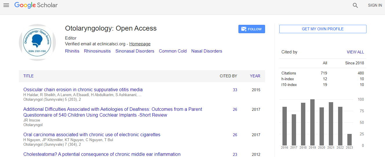
Google Scholar citation report
Citations : 925
Otolaryngology: Open Access received 925 citations as per Google Scholar report
Otolaryngology: Open Access peer review process verified at publons

Indexed In
- Index Copernicus
- Google Scholar
- Sherpa Romeo
- Open J Gate
- Genamics JournalSeek
- RefSeek
- Hamdard University
- EBSCO A-Z
- OCLC- WorldCat
- Publons
- Geneva Foundation for Medical Education and Research
- ICMJE
Useful Links
Recommended Journals
Related Subjects
Share This Page
Otosclerosis as a neoplasm of the outer layer of the otic capsule
Global Summit and Medicare Expo on Head & Neck Surgery
Leslie Michaels and Sava Soucek
The Royal National Throat, Nose and Ear Hospital, UK
Posters-Accepted Abstracts: Otolaryngol (Sunnyvale)
Abstract
We studied stained step sections embedded in celloidin of 54 temporal bones from 27 patients with otosclerosis at the Royal National Throat, Nose and Ear Hospital, London and the House Ear Institute, Los Angeles, California, in order to investigate the histopathologic changes in that disease of the otic capsule (OC). The outer layer of the normal OC is composed of regularly arranged osteons (Volkmann��?s canals and osteocytes) formed from the primitive connective tissue covering the OC externally. Otosclerosis is composed of multiple new plaques in the outer layer of the OC, each showing some resemblance to the structure of the normal outer layer of OC bone. The plaques, however, have each a basophilic area composed of irregular osteons comprising atypical osteoblasts with numerous, irregular Volkmann��?s canals. This has the appearance of an invasive front leading the growth of the otosclerotic plaque. This invasive front is always on the side of the plaque opposite to the periosteal surface of the involved temporal bone. Each plaque also shows increasingly more differentiated and paler-staining bony tissue retrogressively towards the periosteal covering of the OC from which the plaque appears to arise. The invasive front shows infiltration of the tissues that it contacts, by irregularly shaped, thin elongated processes of basophilic osteoblasts. Such invaded tissues are often the inner layer of the OC and beyond this the cochlea and vestibule. Since otosclerotic plaques are most commonly derived from the outer layer of OC near the periosteal connective tissue adjacent to the tensor tympani muscle, the structures most often invaded by otosclerosis are the stapediovestibular joint and the stapes footplate. The presence of multiple, similarly-structured plaques of otosclerosis, each with its undifferentiated invasive front edge and, posterior to that, progressive differentiation of the otosclerotic bony lesion towards the primitive connective tissue base are strongly suggestive of an invasive bony neoplasm of the OC. Otosclerosis has been in its long history rarely considered to be such a lesion. On the basis of the histopathology we put forward here, we consider neoplasia to be the most like pathologic basis for this condition.Biography
Leslie Michaels has completed his Medical education at Westminster Hospital Medical School in London in 1949. He has underwent training in both Clinical and Anatomical Pathology in Bristol, Manchester and London UK. He subsequently practiced Clinical and Anatomical Pathology in North America from 1959, spending most of the time in Northern Ontario, Canada. He has returned to London in 1970 as Pathologist to the Royal National Throat, Nose and Ear Hospital and Professor of ENT Pathology and Dean of the Institute of Laryngology and Otology. His main research interest has been in the pathology of the ear and he has contributed publication in this field. In recent years he has contributed particularly in the field of Meniere’s disease and otosclerosis.
Email: l.michaels@ucl.ac.uk